Do you have a question about the Shenzhen Mindray Bio-Medical Electronics Co., Ltd. DC-N2 and is the answer not in the manual?
| Brand | Shenzhen Mindray Bio-Medical Electronics Co., Ltd. |
|---|---|
| Model | DC-N2 |
| Category | Medical Equipment |
| Language | English |
Classifies equipment safety against electric shock, water ingress, etc.
Defines signal words (DANGER, WARNING, CAUTION, NOTE, Tips) and their meanings.
Explains common safety symbols used in the manual and on equipment.
Provides essential safety guidelines for patient and operator during system use.
Warns about potential allergic reactions to latex in probe sheaths.
Details warning labels attached to the system and their meanings.
Specifies the intended clinical applications and body parts for system use.
States that the system is not intended for ophthalmic use.
Identifies the system's product and model codes.
Outlines the technical specifications including imaging modes and power supply.
Details the standard configuration and available accessories.
Lists compatible peripheral devices like printers and footswitches.
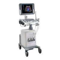
Provides an overview of the system's main components with numbered labels.
Describes the interface panel for input/output signals and ports.
Details the power inlet, auxiliary outlet, and equipotential terminal.
Explains the functions of various buttons, indicators, and knobs on the control panel.
Lists and explains the symbols used throughout the system and manual.
Provides instructions on safely moving and positioning the ultrasound system.
Details how to connect the system to an external power supply or battery.
Describes the correct procedures for powering the system on and off.
Explains how to adjust the monitor's position, tilt, brightness, and contrast.
Provides instructions for safely connecting and disconnecting ultrasound probes.
Guides on connecting the footswitch to the main unit via a USB port.
Details the procedure for safely connecting and removing USB storage devices.
Explains how to connect and set up graph/text printers with the system.
Outlines how to initiate a new patient exam or continue an existing one.
Describes how to enter and manage patient information for exams.
Guides on selecting the appropriate exam mode and probe for scanning.
Explains how to switch between different imaging modes available on the system.
Details on reactivating or continuing previously performed exams.
Provides instructions on pausing and properly ending a patient examination.
Explains how to navigate and switch between different imaging modes.
Covers fundamental image adjustment techniques and parameter controls.
Describes how to save and restore image parameters for quick access.
Details the B mode imaging protocol and optimization techniques.
Explains the M mode protocol and image optimization procedures.
Covers optimization techniques for color Doppler imaging parameters.
Details optimization for Power mode imaging, including gain and map settings.
Explains the PW Doppler mode protocol and image optimization parameters.
Describes the iScape panoramic imaging feature and its procedures.
Covers Smart 3D imaging, its quality conditions, and basic procedures.
Explains splitting display options (dual-split, quad-split) and image magnification.
Details the Spot Zoom and Pan Zoom functions for image magnification.
Describes how to freeze and unfreeze the ultrasound image.
Provides instructions for reviewing captured cine images manually and automatically.
Explains how to compare images within the same exam or across different exams.
Covers cine memory settings, splitting options, and clearing procedures.
Describes how to preset the cine storage length.
Covers entering/exiting measurement modes and understanding results.
Details general measurements for 2D, M, and Doppler modes.
Lists specific measurement types for various clinical applications.
Provides accuracy specifications for different measurement types.
Explains how to add, edit, move, and delete comments on ultrasound images.
Details how to add, move, and delete body marks for anatomical reference.
Covers entering and managing patient information in the system.
Explains storage media, file formats, and saving/reviewing images.
Details how reports are stored, imported, exported, and printed.
Describes the iStation interface for searching, viewing, and managing patient data.
Provides instructions for backing up and erasing files using the DVD drive.
Explains how to view and manage tasks like storage, printing, and media operations.
Details system access settings, user types, and login procedures.
Covers network and DICOM service presets for system configuration.
Explains how to verify the system's network connectivity to DICOM servers.
Details DICOM services like storage, print, worklist, and query/retrieve.
Describes saving patient data to external media in DICOM format.
Covers sending and backing up DICOM structured reports.
Explains how to view and manage DICOM tasks like storage and printing.
Allows presetting various system configurations like region, general, and image.
Enables selection and configuration of probes and exam modes.
Details measurement preset settings, referring to the Advanced Volume.
Covers settings for print services and printer drivers.
Covers local TCP/IP settings and iStorage configuration.
Describes system software updates and other special functions.
Displays product name, hardware version, and software details.
Lists available probes, their illustrations, and basic functions.
Provides essential warnings and procedures for performing biopsy procedures.
Provides an overview of the system's battery and charging process.
Lists important precautions for battery charging and handling.
Explains how to interpret the battery status indicator on the screen.
Recommends performing a full discharge/charge cycle for battery maintenance.
Describes procedures to check the battery's performance over time.
Provides guidelines for the proper disposal of the system's battery.
Discusses potential bioeffects related to diagnostic ultrasound energy.
Emphasizes the importance of using ultrasound prudently for patient benefit.
Explains the ALARA principle for controlling ultrasound energy exposure.
Explains Mechanical Index (MI) and Thermal Index (TI) and their relation to bioeffects.
Covers adjusting acoustic power percentage and default settings.
Details system controls that affect ultrasound output and image quality.
Explains derated ultrasonic output parameters and limits.
Lists measurement uncertainty values for various parameters.
Lists references for acoustic power and safety standards and guidelines.
Provides guidance on electromagnetic emissions compliance for the system.
Details electromagnetic immunity requirements and environments for system use.
Recommends separation distances for RF communication devices to prevent interference.
Outlines daily maintenance tasks for cleaning and checking system components.
Provides a table of common system failures, causes, and solutions.
Describes how to enter and exit the iScanHelper application.
Explains how to use iScanHelper for referencing scanning procedures.
Guides on using iScanHelper for practicing ultrasound scanning techniques.
Details the iScanHelper interface, view selection, and help information.
Notes the unavailability of measurement, comments, and body mark in iScanHelper.
Details inspection criteria for the power cord plug and its components.
Covers visual and contextual inspection of the device enclosure and accessories.
Ensures that device labels and warning labels are present and legible.
Specifies tests for protective earth resistance and acceptable limits.
Describes the procedure and outlet conditions for performing earth leakage tests.
Details the procedure and outlet conditions for enclosure leakage tests.
Covers measurement of patient leakage currents and limits for BF applied parts.
Details tests for mains on applied part leakage and limits for BF applied parts.
Covers measurement of patient auxiliary currents and limits for BF applied parts.
