14
The Eye
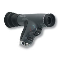
In addition to examination of the fundus, the PanOptic Ophthalmo-
scope is a useful diagnostic aid in studying other ocular structures.
The light beam can be used to illuminate the cornea and the iris for
detecting foreign bodies in the cornea and irregularities of the
pupil.
The practitioner can also easily detect lens opacities by looking at
the pupil through an add-on corneal viewing lens. In the same man-
ner, vitreous opacities can be detected by having the patient look up
and down, to the right and to the left. Any vitreous opacities will be
seen moving across the pupillary area as the eye changes position or
comes back to the primary position.
A) Macula
B) Vitreous humor
C) Sclera
D) Choroid
E) Retina
F) Ora Serrata
G) Canal of Schlemm
H) Anterior chamber
I) Iris
J) Cornea
K) Ciliary body
L) Zonule (Suspensory Ligament)
M) Conjunctiva
N) Lens
O) Hyaloid canal
P) Central retinal vein
Q) Optic nerve
R) Central retinal artery
 Loading...