Do you have a question about the Azure Biosystems Azure 200 and is the answer not in the manual?
Provides warnings and precautions for safe use of UV radiation emitted by the transilluminator.
Details essential safety measures for handling electrical components and avoiding shocks.
Explains the purpose and location of the ground terminal for electrical safety.
Describes the laser illumination system and its compliance with safety standards.
Details safety interlocks and enclosure design to prevent human exposure to radiation.
States that laser systems do not require regular preventive maintenance.
Defines authorized service actions for laser module repair or replacement.
Advises against defeating safety interlock systems for laser imaging operations.
Outlines the warranty terms and conditions for Azure Imaging System products.
Explains that the system automatically selects the correct voltage for the region.
Lists the EC Directives and harmonized standards the system complies with.
Provides contact details for Azure Biosystems, Inc., including email and phone.
Presents detailed technical specifications for the Azure Imaging System models.
Provides contact details for technical assistance regarding installation, setup, or general use.
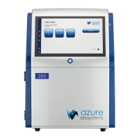
Offers guidelines for optimal placement of the Azure Imaging System in a laboratory environment.
Instructs on how to properly connect the system to a grounded power source, recommending surge protection.
Details the procedure for powering the system on and off, including computer shutdown.
Explains that the acquisition software is pre-installed and how to launch it.
Describes the compatibility and connection of external USB devices like keyboards and mice.
Introduces the main imaging options available on the software's home page.
Explains essential imaging parameters like Auto vs Manual, Sensitivity, Exposure, and Live Mode.
Details the step-by-step process for capturing chemiluminescent images.
Guides users through capturing fluorescent images with multiplexing capabilities.
Provides steps for imaging nucleic acid blots using various channels.
Outlines the procedure for capturing protein gel images.
Describes how to create and customize imaging protocols for specific needs.
Explains how to access, open, save, and close images within the gallery.
Details image analysis tools like adjustments, ROI, transform, annotations, and info.
Covers selecting regions of interest and applying image transformations like rotation and flipping.
Describes adding annotations and viewing detailed image acquisition parameters.
Covers dye lists, file saving, simulation mode, and system preferences.
Explains creating darkmasters and performing focus calibration.
Details managing user accounts and changing system passwords.
| Detection Method | CCD Camera |
|---|---|
| Software | AzureSpot Analysis Software |
| Applications | Protein Quantification |
| Maximum Sample Size | 20 x 20 cm |
| Sample Format | Gels, Blots |
| Computer Interface | USB |