Do you have a question about the Instrumentarium ORTHOPANTOMOGRAPH OP300 and is the answer not in the manual?
Scan patient with open bite, install positioning plate, take scout image, and select scan parameters.
Begin the dental planning process using specialized drill guide planning software.
Scan patient with radiographic guide, install positioning plate, and configure scan settings.
Commence the dental planning process with the drill guide planning software.
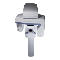
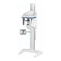
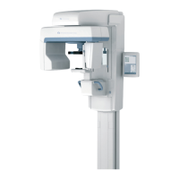
| Model | ORTHOPANTOMOGRAPH OP300 |
|---|---|
| Focal Spot Size | 0.5 mm |
| Type | Panoramic X-ray |
| Manufacturer | Instrumentarium |
| Imaging Modalities | Panoramic, Cephalometric |
| X-ray Source | X-ray tube |
| Image Receptor | Digital sensor |
| Detector Type | CCD or CMOS |
| X-ray Tube Voltage | 60-90 kV |
| X-ray Tube Current | 4-16 mA |
| Patient Positioning | Standing or sitting |
| Image Area | Panoramic: Up to 15 x 30 cm |
| Power Requirements | 230 VAC, 50/60 Hz |
| Field of View | Panoramic: 150 x 300 mm, Cephalometric: 240 x 300 mm |