Do you have a question about the Konica Minolta mKDR Xpress and is the answer not in the manual?
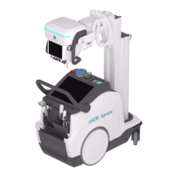
Details the main characteristics of the mobile X-ray unit, including design, power, and controls.
Explains the information found on identification labels attached to the equipment.
Specifies the intended use, normal operation, and contraindications for the X-ray system.
Lists the parts of the X-ray equipment that come into physical contact with the patient.
Crucial safety instructions for operators and service personnel, emphasizing manual study.
Defines liability and responsibilities for safe operation, maintenance, and modifications.
Explains radiation dose limits and safe practices, emphasizing ALARA principle.
Details measures to minimize radiation exposure, including lead shielding and distance.
Defines various safety symbols used throughout the manual and their meanings.
Covers compliance with safety standards (IEC, CFR) and certifications for the unit.
Provides compliance information for RF and harmonic emissions according to IEC standards.
Details immunity test levels and compliance for various environmental factors like ESD and surges.
Describes performed tests and provides quantitative data for image performance.
Lists main controls like Control Panel, Console Screen, Handswitch, and Collimator.
Describes controls for unit movement and arm positioning, including safety.
Details how to connect the unit to the power supply and safety requirements.
Describes the primary display and Konica-Minolta Ultra software application.
Explains the displays and information shown on the head-assembly console.
Explains the meaning of different indicator colors during system operation.
Details methods to turn the unit ON using RFID or Keypad systems.
Step-by-step guide for powering ON using RFID cards and reader.
Step-by-step guide for powering ON using the ON/OFF button and keypad.
Instructions for safely turning the unit OFF using the ON/OFF button.
Explains how to use the emergency stop switch and its safety implications.
Describes the sequence of events when turning the system ON and OFF.
Explains how battery status is displayed for X-ray and motion functions.
Details system alerts for low battery conditions and recommended actions.
Describes settings for automatic shutdown and inactivity timeouts.
Explains the meaning of system status indications displayed by the light.
Details available ports for peripherals, network connectivity, and detector cables.
Explains the operation and indicators of the handswitch for exposures (Prep/Exp).
Details the functionality and usage of the optional remote control for exposures.
Emphasizes safety during unit movement: parking, inclines, surfaces, and collisions.
Explains how to drive the unit using the handlebar and its height adjustment.
Describes controls for precise system positioning and safety measures to prevent movement.
Details how to disengage motors for manual unit movement and associated safety.
Explains the function of proximity sensors in reducing speed and alerting users.
Describes bumper sensors that stop motor movement upon frontal collision.
Explains the option to release the driving brake in emergency or breakdown situations.
Instructions on how to place the arm in its parking position for safety and stability.
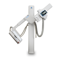
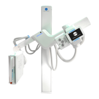
Details controls for rotating the column and moving the telescopic arm.
Explains how to rotate the head-assembly on its transversal and horizontal axes.
Describes the rotation of the collimator on its vertical axis and required SID.
Covers filtration options, laser, radiation meter, and DAP chamber.
Describes the sensor for measuring focus-to-patient distance and its display.
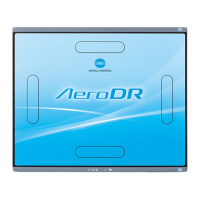
Introduces detector models and their installation with handles and grids.
Explains how detectors are stored and charged in unit holders and power supply.
Describes the cable for wired detector configuration and storage.
Details the barcode scanner for patient information registration.
Provides guidance on handling, cleaning, and environmental considerations for detectors.
Describes the primary display and Konica-Minolta Ultra software application.
Explains the displays and information shown on the head-assembly console.
Explains the meaning of different icons indicating system status during exposure.
Describes how the heat unit calculator displays tube thermal capacity usage.
Details the display of battery charge levels in various modes (charging, stand-alone).
Explains indicators for unit parking status and movement speed (turtle/rabbit).
Covers mute button, user prompts, and workstation selection interface.
Explains how to select kV, mA, mAs, and Time for exposures.
Details the use of the slider for increasing/decreasing technique values.
Covers focal spot selection, power limitation, and head-assembly rotation views.
Explains how to view focal-skin distance and select collimator filters.
Details the location and function of the message bar on the console.
Explains different pop-up message windows and their content (notifications, errors).
Describes how to generate and export system event logs for troubleshooting.
Categorizes messages into Warning, Information, Inhibit, User Action, and Error.
Lists specific error codes, their descriptions, and recommended user actions or service calls.
Provides a procedure for warming up the X-ray tube to preserve its life.
Details the typical sequence for performing a radiographic examination.
Instructions for aligning the X-ray beam with the patient and receptor.
Guidelines for maintaining battery health, including charging and storage.
Tasks to be performed for periodic maintenance, including charging and cable checks.
Instructions for safely cleaning and disinfecting the unit's external surfaces.
Encompasses power, kVp, mAs, mA, time, focal spot, duty cycle, and tube specs.
Details power line, battery capacity, output accuracy, heat, and environmental conditions.
Provides details on the E7886 X-ray tube insert, including focal spots and filtration.
Lists the physical dimensions (length, width, height) and weight of the unit.
Emphasizes careful positioning and use of immobilizing devices for children.
Covers shielding, minimizing dose, and adjusting technique factors for pediatric exams.
Recommends antivirus software and limiting system access to authorized users.
Outlines practices for ensuring software integrity and data security.
Explains how to access the administrator settings utility via the system status icon.
Describes available settings like Operator Settings and Maintenance.
Covers language, units, time zones, sound, visual, and power-off configurations.
Details how to check system information, logs, and export data.
Explains how to manage RFID tags for system access and permissions.
| A/D Conversion | 16-bit |
|---|---|
| Spatial Resolution | 3.6 lp/mm |
| Power Requirements | 100-240 VAC, 50/60 Hz |
| Type | Digital Radiography System |
| Detector Size | 17" x 17" (43 cm x 43 cm) |
| Pixel Pitch | 139 μm |
| X-ray Generator | High Frequency Generator |
| Voltage Range | 40-125 kV |
| Exposure Time | 1 ms |
| SID Range | 100-180 cm |
| Interface | DICOM 3.0 |