
V
Vanessa DunnAug 2, 2025
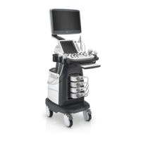
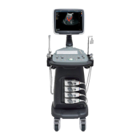
How to troubleshoot if the MediSono Medical Equipment ultrasound system cannot be started up?
- EEric ChapmanAug 2, 2025
If the MediSono Medical Equipment ultrasound system cannot be started, verify that it is plugged in and that the main power switch is in the correct position. Also, check the fuse to confirm it is intact. If the fuse is blown, replace it.