Do you have a question about the MediSono P25 EXP and is the answer not in the manual?
Defines the system's intended applications and limitations.
Details electrical safety measures to prevent shock and equipment damage.
Outlines safe practices for moving and handling the system to prevent injury.
Addresses safety measures related to biological hazards and infection control.
Explains acoustic power principles, including biological and thermal indices.
Identifies and explains important safety symbols used on the system.
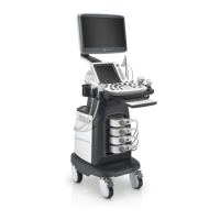
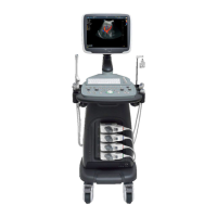
Lists the main components included in the standard system configuration.
Provides physical dimensions and weight of the ultrasound system.
Details the various physical components of the ultrasound system.
Describes the ports and connectors on the peripheral device panel.
Explains the layout and functions of the control panel buttons and knobs.
Identifies and explains the elements displayed on the system's basic screen.
Details the functionality and controls available on the touch screen interface.
Provides instructions for assembling the main system components.
Guides on how to safely move and position the ultrasound system.
Explains how to connect the system to power sources.
Steps for connecting the ultrasound probe to the system.
Details on connecting external devices like printers and footswitches.
Allows configuration of general system parameters like language and date.
Covers settings related to image display formats and screen savers.
Configures settings for saving images and cine clips.
Allows customization of function keys for quick access to features.
Configures measurement parameters, formulas, and lists for applications.
Configures system settings for DICOM connectivity and services.
Displays current hardware, software, and touch screen information.
Guides on how to calibrate the touch screen for accurate input.
Steps for entering and managing patient information before an exam.
Procedures for pausing and resuming an ongoing patient examination.
Guidance on how to complete or discontinue a patient examination session.
Guides on selecting the appropriate probe and exam type for imaging.
Steps to enter and optimize B-mode imaging for anatomical structures.
Details CFM, PDI, and TDI modes for color flow imaging.
Steps to acquire and display M-mode images for cardiac applications.
Procedure for acquiring Pulsed Wave (PW) and Continuous Wave (CW) Doppler images.
Guidance on adjusting parameters for color flow, PDI, and TDI image optimization.
Guidance on adjusting parameters for spectral Doppler image optimization.
Provides guidelines for the correct usage, cleaning, and sterilization of ultrasound probes.
Details on biopsy bracket assembly, preparation, verification, and performance.
Step-by-step instructions for cleaning ultrasound probes.
Details the process for disinfecting and sterilizing ultrasound probes.
Provides instructions for assembling biopsy brackets for probes.
Guidance on performing a biopsy using the system's features.
Procedures for cleaning the system's external surfaces, dust filter, and trackball.
Lists essential maintenance checks to be performed annually.
Provides solutions for common system issues and error messages.
Instructions for safely replacing system fuses to restore power.
Details the system's compliance with various safety and electrical standards.
Information on the system's electromagnetic emissions and environment.
Details the system's immunity to electromagnetic interference.
Provides recommended separation distances to prevent RF interference.
Lists compatible coupling gels for different probe models.
Lists compatible cleaners for different probe models.
Lists compatible disinfectants for different probe models.
| Brand | MediSono |
|---|---|
| Model | P25 EXP |
| Category | Medical Equipment |
| Language | English |