Hear more, Live better!
03 04
Operation Manual of Cochlear Implant
08
Measuring Cochlea size and selecting electrode length
The cochlea implanting operation shall be performed in accordance with the Operating Guidance
on Implantation of Cochlea published by China Medical Association, which mainly includes
following procedures:
1) Make an incision behind the ear, detach skin flap, cut open musculo-periosteal flap, and expose
the mastoid and back wall of bony external auditory meatus;
2) Open the mastoid cavity;
3) Grind a bone bed of 15mm in diameter and 2.5mm in depth on the surface of skull;
4) Open facial recess and perform fenestration operation of the cochlea;
5) Place implant in the bone bed and implant electrode in the tympanic canal;
6) Stitch in sequence musculo-periosteal flap, subcutaneous tissue and skin;
7) Carry out impedance test of implant during the operation.
Notes:
1) The implanting operation of cochlea shall be performed by professional doctors who are skillful
in otology micro-surgery and have taken specialized trainings on cochlea operations.
2) Maintain the surface facing the cochlea axis and insert the electrode in, until the marking on the
end of electrode contact array reaches the opening of cochlea.
3) If impedance test of implant is to be conducted before stitching skin flat, temporarily restore
skin flap and / or use other means to enhance the contact between implant and tissues, so as to
decrease the impedance of implant. Operating distance of transmission coil of speech processor is
3-10mm. Beyond this distance, the system may not function properly.
10
Operative Complications
Cochlear implantation is a relatively safe operation, with such commonly seen complications as:
Common complications: subcutaneous hematoma; tympanum or external auditory meatus
perforation; facial nerve or tympanic cord nerve paralysis; implant is prolapsed if it is not
implanted to a sufficient depth and wearer's skull is too thin, or signal transmission is affected if
the skull is thick; dizziness; tinnitus; facial tic or pain during electric stimulus. Severe
complications: Electrode prolapse or implant displacement; severe infections of incision/
breakdown; necrosis of skin flap; massive haemorrhage due to damage of emissarium mastoideum
or sigmoid sinus; leakage of cerebrospinal; facial nerve paralysis; electrode failure; taste
disturbances.
Other complications: Implanted silicone allergy; Device failure; Wound Swelling; Wound
breakdown
Declining cognitive function; Acute otitis media; Pain; Non-auditory sensation.
The product is applicable to severe-to-profound sensorineural deafened patients aged 12 months
and above. Implant is the implantable part of the cochlear system, used with the NSP-60C speech
processor produced by Nurotron Company. The part has been certified by national or local SFDA
departments. Its commissioning shall be conducted with the aid of NuroSound fitting software.
Absolute contraindications:
1) Serious malformation of inner ear, such as Michel deformation or cochlea absence;
2) Auditory nerve absence;
3) Deafness caused by non-cochlea illness;
4) Serious mental disease;
5) Uncured purulent mastoiditis of middle ear.
Relative contraindications:
1) Bad physical condition;
2) Uncontrollable epilepsy.
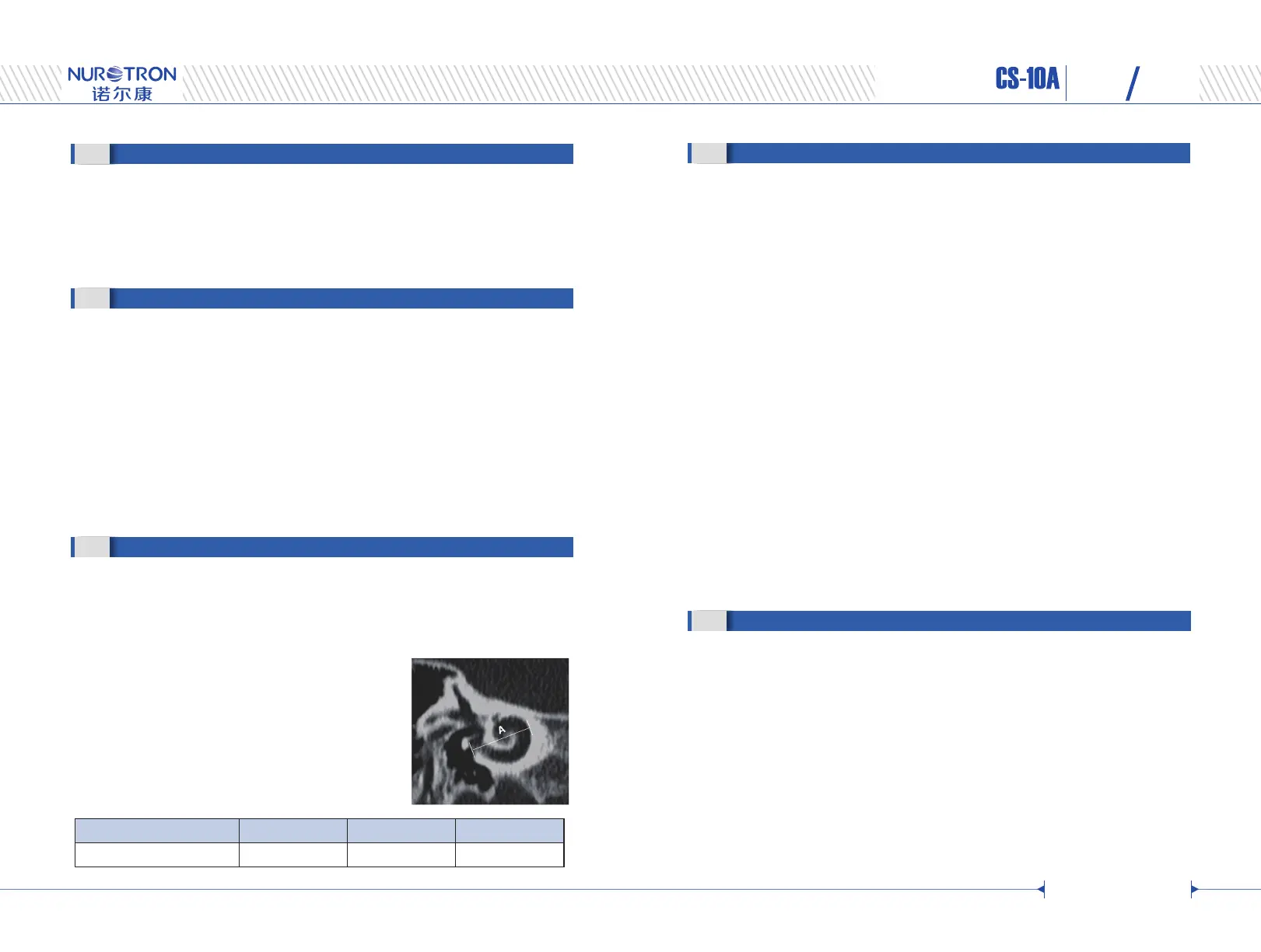
CS-10A cochlear implant electrodes have three different lengths: TS 16mm, TM and SD 20.5mm
and TL 24mm. To maintain a relatively constant insertion depth for patients with different
cochlear sizes, a small cochlea shall fit a short electrode and a large cochlea shall fit a long array.
The cochlea size shall be estimated by measuring the basal diameter of the cochlea (defined as A
value as shown in the figure below) on a CT scan image with
an oblique coronal view or the axial plane view. The A value
is measured from the mid-round window through the center
of the cochlea to the lateral wall. Based on the A value,
electrode type can be selected per the Table below.
Other methods to estimate cochlea size can be also used at the
surgeons’ discretion.
< 8 mm 8 – 10 mm > 10 mm
TS electrode SD /TM electrode TL electrode
Cochlea Diameter A value
Electrode Type
 Loading...