17www.sam-dental.de
Bergstrom G.
On the reproduction of dental
articulation by means of articulators,
a kinematic investigation.
Acta Odontol Scan 1950;9 (suppl
4):125-141
Baldauf A, Mack H, Wirth CG.
Bestimmung der Scharnierachse
mittels des äußeren Gehörgangs.
Info Orthod Kieferoothop
1996;28:459-465
Nagy WW, Smithy TJ, Wirth CG.
Accuracy of a predetermined
transverse horizontal mandibular axis
point. J Prosthet Dent
2002;87:387-394
Es wurde eine Studie durchgeführt, um die Beziehung der
Mitte der sagittalen und horizontalen Ebene bezüglich der
anthropologischen knöchernen Gehörgänge zu bestimmen.
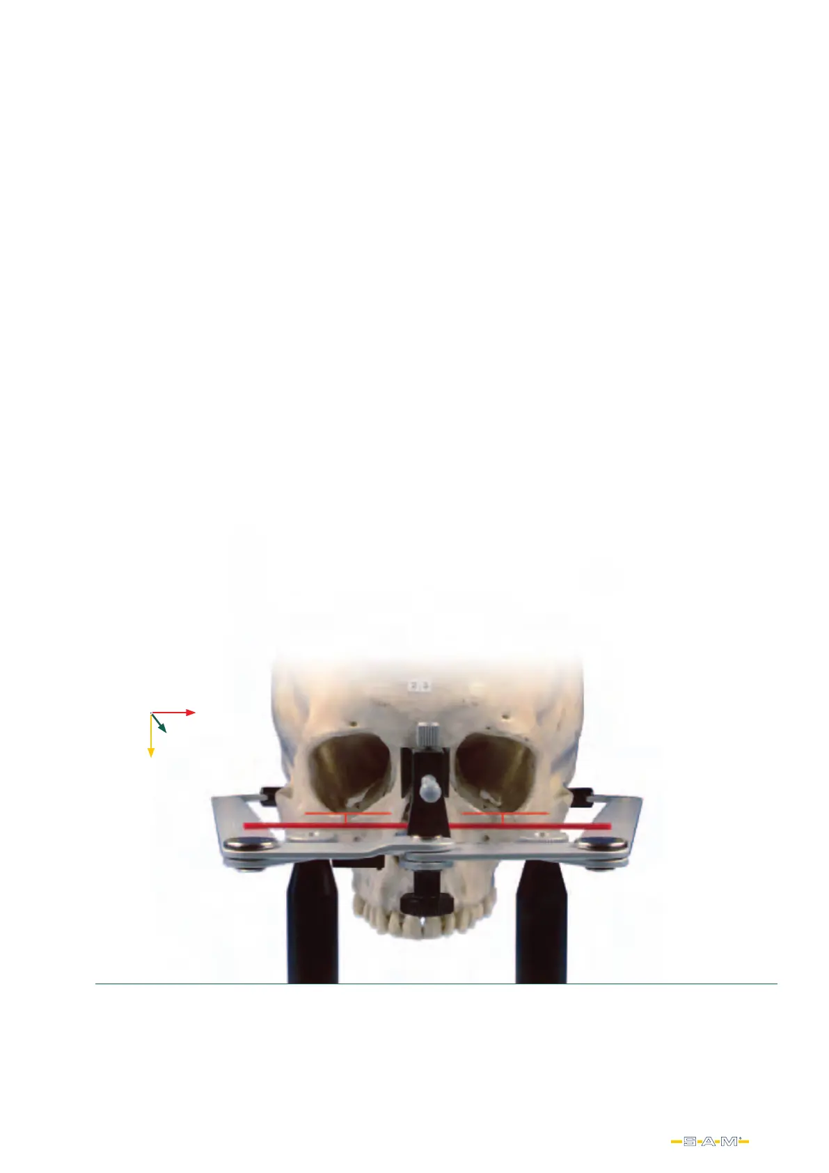
Der SAM® AXIOQUICK® Gesichtbogen wurde auf einen
speziellen Montagestand befestigen. Dieser Bogen wurde
auf 122 Schädel befestigt und digital fotografiert. Es
konnte hierbei festgestellt werden, dass die Untergrenze
der Orbitale stets mit der Horizontal ebene des Bogens
ausreichend parallel waren. Ein gleiches Ergebnis wurde
an weiteren 44 Patienten erzielt, d. h. die Horizontalebene
war stets mit der Interpupillarebene ausreichend parallel.
Die Vermessung mittels Photoshop 6.0 ergab in 70% der
Fälle Seitendifferenzen der Schädel, wie auch Patienten
von ≤ 1 mm und Seitenabweichungen von < 2 mm. Diese
Studie zeigt, dass ein richtig konstruierter Transferbogen
bei entsprechender Handhabung die schädelgerechte
Übertragung des Oberkiefermodells in den Artikulator
gewährleistet.
Warum diese Anlegetechnik und nicht anders?
Why this alignment technique should be used.
X
Z
Y
A study was done to determine the mid-sagittal and
horizontal plane relationships in reference to the
anthropological bony ear canals. The SAM® AXIOQUICK®
facebow was modified to attach to a special mounting
stand This facebow was attached to 122 skulls and digitally
photographed. It could be noted that the lower limit of the
orbital eye sockets were always sufficiently parallel with
the horizontal plane of the facebow. A similar result was
obtained in another 44 patients, ie, the horizontal plane
was always sufficiently parallel with the Interpupillary plane.
The measurement using Photoshop 6.0 was found in 70%
of cases side differences of the skull, as well as patients of
≤ 1 mm and lateral deviations of <2 mm. This study shows
that a properly designed facebow with appropriate handling
ensures a correct skull releated transfer of the maxillary
model to the articulator.
Ergebnisse / Results:
Sowohl in der Gruppe der „historischen“ Schädel, als
auch in der Gruppe der Patienten sind ca. 70% der
gemessenen Seitendifferenzen ≤ 1 mm. In den größeren
Abweichungsbereichen beobachtet man jedoch eine höhere
Variabilität bei den Schädeln. In der Gruppe der Patienten
waren alle Seitendifferenzen ≤ 2 mm, in der Gruppe der
Schädel waren 8% > 2 mm. Der Unterschied der Streuung
der Messwerte wird durch den Variationskoeffizienten
bestätigt.
Both in the group of „historic“ skulls, as well as in the group
of patients are approximately 70% of the measured side
differences ≤ 1 mm. In the larger deviation areas, however,
we observed a higher variability in the skulls. In the group
of patients were all side differences ≤ 2 mm, in the group of
the skull were 8%> 2 mm. The difference in the scattering
of the measured values is confirmed by the coefficient of
variation.
 Loading...