30 2210200496L41 1904V004
Usage
Sinus image acquisition
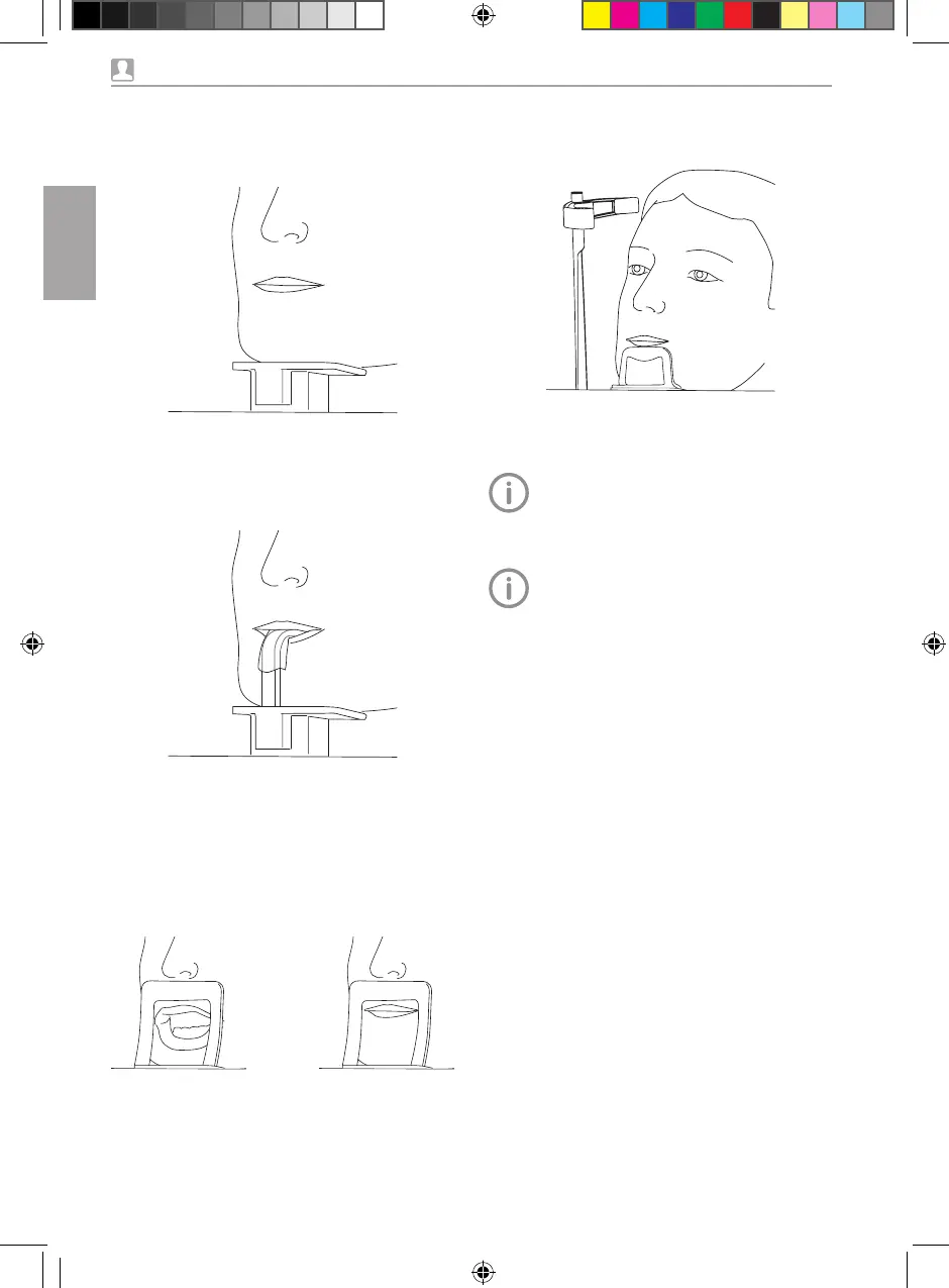
i Position the patient so that his/her bottom lip
presses lightly against the chin support�
Adjusting the position with the positioning
beams
The positioning beams use class 1 laser
beams� Although these can dazzle the
patient, they are safe and will not dam-
age the eye�
For CBCT images it is sufficient to do the
positioning based on the mid-sagittal
plane and the Frankfurt plane�
For all other image types the patient
needs to be positioned more accurately
with the aid of the following steps�
The alignment of the upper canine X-ray
positioning beam is crucial for the image
quality of panoramic images�
i Check to make sure that the patient has
closed his/her eyes�
i If necessary, correct the height of the unit
again�
i Activate the positioning beams with the but-
ton�
CBCT image acquisition
i The patient places his/her chin on the mount-
ing for the bite block�
The bite block is not required�
Panoramic image acquisition
i The patient bites onto the bite block, with the
upper and lower incisors resting in the
grooves provided�
i Use the chin support for edentulous patients
in the case of patients who do not have any
teeth� Here, the patient places his/her chin on
the chin support�
Maxillary joint image
i Position the patient with the upper lip against
the chin support�
EN
US
OBJ_DOKU-0000003575-mehrere-005.indd 30 17.04.2019 14:29:01


 To Next Page