(4D Views TEE – cont.)
or use the rotary knobs from the scanner assigned respectively for control
of the data
ME alignment
A triplane image and a short axis view appear on the screen
1. The long axis of the ventricle needs to be aligned with the center red line
2. LV axis should be straight down in the middle of the image
3. The blue line needs to be positioned on the MV annulus
4. Use the trackball or mouse to
control the datasets
Use the
to move and to
position the data set
Use the to tilt the views
Grab the blue lines and move it to the MV annulus
5. Once alignment is done press Approve button
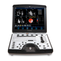
Vivid E9 EchoPAC
4D Views

 Loading...