Do you have a question about the GE Vivid S70 and is the answer not in the manual?
Provides general information about the manual and its contents, including safety precautions and documentation structure.
Details the various manuals comprising the Vivid S70 / S60 documentation set and their purpose.
Specifies the intended uses and applications for the Vivid S70 / S60 system.
Lists conditions or uses for which the Vivid S70 / S60 system is not intended.
Lists GE Ultrasound contact details for information, service, and placing orders in various regions.
Explains different levels of safety precautions identified by DANGER, WARNING, and CAUTION icons.
Outlines the owner's responsibility to ensure proper system operation, maintenance, and operator training.
Defines thermal index (TI) and mechanical index (MI) parameters related to ultrasound safety.
Details crucial safety aspects including patient safety, personnel safety, and environmental protection.
Covers essential site requirements including power, environment, and electromagnetic interference considerations.
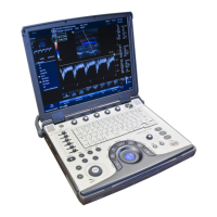
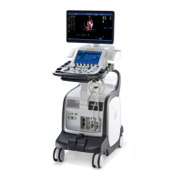
Introduces the system's components and layout, including the front and rear views of the Vivid S70 / S60.
Provides instructions on how to connect and disconnect probes, including safety precautions.
Details the layout and functions of the system's control panel, including key illumination and adjustments.
Explains procedures for safely moving and transporting the ultrasound system, including wheel operations and precautions.
Guides users through creating new patient records and selecting existing ones before starting an examination.
Explains how to use the cineloop feature to review and store image sequences, including marker adjustments.
Describes the two types of zoom functionality: Display zoom and High resolution (HR) zoom.
Describes how to insert, edit, and manage text and bodymark annotations on images.
Details the overview and optimization techniques for the 2D imaging mode, including gain and TGC controls.
Explains the different types of M-Mode and how to use and optimize them for measurements.
Covers the overview and optimization of Color 2D and Color M-Mode, including adjustments for flow display.
Describes how to use and optimize PW/CW Doppler modes for spectral analysis and flow assessment.
Explains the overview and optimization of Tissue Velocity Imaging for assessing tissue velocities.
Details the overview and optimization of Tissue Tracking for analyzing myocardial displacement.
Explains the overview and optimization of Strain rate calculations for tissue deformation analysis.
Introduces the 4D imaging capabilities, including real-time single and multi-beat acquisition.
Explains the Bi-plane and Tri-plane modes for viewing multiple scan planes simultaneously.
Describes FlexiViews for quick access to standard or user-defined views in 4D TTE or TEE scanning.
Provides an overview of the integrated stress echo package and its capabilities for image acquisition, optimization, and analysis.
Guides users on how to select or create stress test protocol templates for examinations.
Describes the process of acquiring images according to a selected template, including continuous capture.
Explains how to view saved loops, assign scores, and perform wall motion analysis.
Details the use of Quantitative TVI for stress echo analysis, including Vpeak and Tissue Tracking.
Covers the process of creating and editing custom stress echo protocol templates.
Explains how to use the Assign and Measure functions for pre-labeled measurements.
Describes the Measure and Assign process for generic measurements and how to apply labels.
Covers advanced measurements like event timing, TSI, AFI, and AutoEF for cardiac analysis.
Details analysis tools for 4D/multi-plane Left Ventricle (LV) quantification.
Describes the 4D Auto MVQ tool for automated mitral valve quantification.
Explains the 4D Auto AVQ tool for automated aortic valve quantification.
Covers measurements for vascular applications like Intima-Media Thickness (IMT).
Details how to use OB graphs and fetal trending for assessing fetal growth and patient data.
Explains how to view, minimize, move, and delete measurement results.
Describes the function of the worksheet for reviewing, editing, deleting, and printing measurements.
Discusses factors affecting measurement accuracy, including image quality, operator variability, and algorithms.
Introduces the Q Analysis software package for analyzing TVI, Strain, Strain rate, TSI, and Contrast data.
Provides instructions on generating traces, manual tracking, and optimizing sample areas within Q Analysis.
Explains how dataflows enable communication and transfer of patient information and images between system components.
Details how to store images and cineloops to the system, removable media, or network folders.
Covers searching, opening, editing, and deleting patient records and examinations from the archive.
Describes communication and connection options for networking and data management with other devices.
Explains how to manage hard disk space by moving, copying, or deleting files and configuring auto-purge.
Outlines procedures for backing up and restoring patient archives and system configurations.
Introduces Tricefy, an online platform for sharing and archiving medical imaging data.
Explains the system's capability to stream live ultrasound data over a network connection.
Describes the system's ability to create patient and examination reports using templates and custom designs.
Details how to create reports by adding data, images, and text, and how to manage other information.
Explains how to open, choose templates, change patient information, add images, print, and store reports.
Introduces the feature for inserting pre-configured diagnostic statements and codes into patient reports.
Describes how to insert comments and get an overview of completed measurements in the final report.
Covers creating and designing custom report templates using information containers.
Explains configuration, deletion, and export/import of user-defined report templates.
Details how to customize global and application-specific settings, system connectivity, and data management.
Explains how to configure the shortcut bar on the touch panel for quick access to functions.
Covers operator registration, user groups, rights, and security settings like auto logon and storage encryption.
Describes how to configure imaging settings such as cineloop store, crop images, Doppler, and patient info display.
Covers basic operations, configuration of measurement menus, user-defined formulas, and measurement accuracy.
Explains system setup for network connectivity, TCP/IP settings, DICOM configuration, and wireless network setup.
Details configuration of archiving functions, patient list management, and dataflow adjustments.
Describes how to build and import customized protocols for automated examinations.
Covers configuring application lists per probe, renaming, deleting, and arranging presets.
Details configuration of TEE probe handle buttons for live and freeze modes.
Lists supported probes, their modes, and technical data including frequency, footprint, and FOV.
Shows a table of available probe presets for various clinical applications.
Provides instructions for general probe care, cleaning, disinfection, and inspection.
Details electrical, mechanical, and biological hazards associated with probe use and handling precautions.
Explains biopsy capability for probes, required guidance systems, and precautions for biopsy procedures.
Introduces peripherals that can operate with the ultrasound system, including printers and streaming boxes.
Explains how to print images using the P1 key and thermal video printer setup.
Covers configuring the P1 button for various actions like printing and DICOM storage.
Details how to configure the system for external monitors, including resolution and EDID settings.
Explains how to connect and configure external monitors for video output, including image quality considerations.
Describes the View-X option for streaming external video signals and its setup via TCP/IP.
Explains interfacing with the CartoSound system for 3D electroanatomical navigation and ultrasound catheter use.
Provides guidance on system maintenance, expected service life, and inspection procedures.
Describes how to handle system malfunctions, bookmark issues, and generate log files.
Explains the process for downloading and installing software updates, including backup procedures.
Details how to save TCP/IP settings to retain them after a software upgrade.
| Power Requirements | 100-240 V AC, 50/60 Hz |
|---|---|
| Application | Cardiac, Vascular, Abdominal, OB/GYN, Small Parts, Pediatric |
| Operating Modes | Color Doppler, PW Doppler, CW Doppler, Tissue Doppler Imaging |
| Advanced Imaging Options | AutoEF |
| Connectivity | DICOM, USB, Ethernet, HDMI |
| Transducer Ports | 4 |
| Weight | Approximately 80 kg |
| Transducer Frequency Range | 1.5 MHz |
| Operating Temperature | 10°C to 40°C |
| Storage Temperature | -20°C to 55°C |
| Humidity Range | 30% to 80% non-condensing |
| Image Modes | B-Mode, M-Mode, Color Doppler, Power Doppler, Pulsed Wave Doppler, Continuous Wave Doppler, Tissue Doppler |
| Supported Probes | Linear, Convex, Phased Array, Volume Probes |
| Probe Connectors | Compatible with a wide range of probes |
| Measurement Tools | Comprehensive cardiac measurement package, including LV, RV, LA, RA, valves, and great vessels |