Do you have a question about the Gendex GXS-700 and is the answer not in the manual?
Describes the sensor's ability to capture diagnostic X-ray images and patient safety.
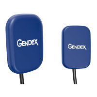
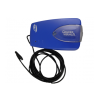
Defines the GXS-700 as a USB digital sensor for acquiring dental intraoral radiography images.
Details the GXS-700 as a CMOS sensor connected via USB for dental imaging.
Advises users on learning to use the GXS-700 sensor effectively and recommends training.
Explains the symbols and conventions used in the manual to convey important information.
Provides instructions on how to unpack and identify GXS-700 system components from the shipping container.
Illustrates the contents of the GXS-700 system package, categorized by tier.
Lists and describes the main system components, including the digital intraoral sensor and USB connector.
Details the various positioning devices designed to support and align the GXS-700 sensor with the X-ray beam.
Explains the use of disposable sheaths as a barrier for the sensor and their packaging.
Describes the USB extender cable included with the GXS-700 system.
Describes the sensor cradle as a solution for safe and secure storage of GXS-700 sensors.
Lists the software and calibration files included on CD-ROMs with the GXS-700 system.
Lists the documentation provided with the GXS-700 system, including user manuals.
Instructs users to always inspect the sensor, cable, and positioning devices for physical damage before use.
Outlines the electrical safety requirements for using the GXS-700 sensor and its components, conforming to IEC standards.
Explains that mains isolation occurs at the computer's input and the sensor can be disconnected.
Reminds users to adhere to dental radiography rules and use protection for patients during X-ray exposure.
Advises on using new hygienic barriers for each patient to prevent cross-contamination.
Provides information on the lead content of the sensor and advises contacting the dealer for disposal information.
Instructs on proper disposal of sheaths and consumables as biomedical waste to prevent disease spread.
Lists minimum and recommended system requirements for the PC to operate the GXS-700.
Recommends frequent backup of patient and image data onto removable storage devices for data recovery.
Mentions that other imaging software may have different requirements and stresses the importance of intended use.
Provides a step-by-step guide for installing the GxPicture software and USB driver for the GXS-700.
Details how to install the sensor cradle, ensuring convenient placement and safety.
Explains the meaning of different status icons displayed in the Windows System Tray, indicating sensor status.
Lists the options available by right-clicking the GXS-700 Status Icon: Information, Image Settings, and Service.
Describes the information dialog that displays connected sensors, friendly names, and version details.
Explains how to verify recognition and communication when multiple GXS-700 sensors are installed.
Details how to adjust image parameters like Gamma, Brightness, Contrast, and Optimizer settings within GxPicture.
Explains the functions available in the Service dialog, such as changing event log location and generating test images.
Guides users through the process of acquiring radiographic images using the GXS-700 and imaging software.
Provides instructions on correctly positioning the sensor in the patient's mouth for X-ray imaging.
Describes alternative methods for positioning the sensor, including manual holding by the patient.
Explains the ease of moving and connecting the GXS-700 between different dental chairs using USB technology.
Provides crucial CAUTIONary advice on removing sheaths, cleaning the sensor, and disinfecting holders after each patient.
Lists recommended cleaning agents and solutions for disinfecting the sensor and holders, with a CAUTION against autoclaving.
States that the GXS-700 requires no special maintenance beyond regular cleaning and disinfection.
Outlines factors affecting image quality and recommends periodic review.
Discusses appropriate dose and exposure time settings for optimal image quality and patient safety.
Explains factors influencing image sharpness and contrast and recommends phantom use for assessment.
Refers to the software manual for guidance on optimal display settings and monitor properties.
Lists detailed technical specifications of the GXS-700 sensor, including dimensions, resolution, and software support.
Details the environmental conditions for usage, transportation, and storage of the GXS-700 sensor.
Presents data on Detective Quantum Efficiency versus spatial frequency as a function of X-ray dose.
Explains various symbols used on the product and accompanying documents, including safety and certification marks.
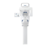
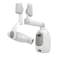
Discusses the compatibility of the GXS-700 with different dental X-ray units and generators.
Explains the correlation between X-ray source distance and the dose received by the sensor.
Lists the software requirements for using the GXS-700, including GxPicture and compatible imaging software.
Provides the EC Declaration of Conformity, listing relevant standards and directives.
Lists the harmonized standards under which conformity is declared, relating to safety and performance.
Offers troubleshooting steps for when the system fails to acquire X-ray images.
Lists various accessories available for the GXS-700, including sheaths, cables, and positioners, with their codes.
Details specific positioner kits, anterior, posterior, bitewing, and endo holders for the GXS-700.
Provides guidance on electromagnetic emissions from the GXS-700 sensor and its compliance with standards.
Outlines electromagnetic immunity tests and compliance levels for the GXS-700 sensor.
Advises on recommended separation distances between RF communication equipment and the GXS-700 sensor.
| Sensor Type | CMOS |
|---|---|
| Connectivity | USB 2.0 |
| Cable Length | 2.5 meters |
| Sensor Sizes | Size 1, Size 2 |
| Image Resolution | 20 lp/mm |
| Compatibility | Windows |
| Resolution | 20 lp/mm |
| Gray Scale | 16-bit |