Do you have a question about the HEIDELBERG Spectralis and is the answer not in the manual?
Remove objective lens, use air bulb for dust, clean with dry microfiber cloth in circular movements.
Use isopropyl alcohol (70%) or ethanol (80%) for disinfection. Follow guidelines for contact times and check for remnants.
Use a moist, well-wrung microfiber cloth with standard cleaning products (no acetone/hydrogen peroxide).
Use recommended disinfectants (e.g., CaviWipes, Descosept AF) suitable for plastic and metal surfaces.
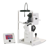
The SPECTRALIS is an advanced ophthalmic imaging device designed for detailed examination of the eye. It is a sophisticated piece of equipment used in clinical settings to capture high-resolution images, aiding in the diagnosis and management of various ocular conditions. The device is engineered to provide precise and reliable results, making it a valuable tool for eye care professionals.
The primary function of the SPECTRALIS is to acquire optical images of the eye, specifically focusing on the retina and other posterior segments. It utilizes advanced optical coherence tomography (OCT) technology to generate cross-sectional views of ocular structures, allowing for detailed visualization of tissue layers that are not visible with traditional ophthalmoscopy. This capability is crucial for detecting subtle changes indicative of diseases such as glaucoma, macular degeneration, and diabetic retinopathy. Beyond OCT, the SPECTRALIS can also perform other imaging modalities, including fundus photography and angiography, providing a comprehensive suite of diagnostic tools. The integration of multiple imaging techniques within a single platform enhances diagnostic accuracy and streamlines the examination process. The device is designed to capture images quickly and efficiently, minimizing patient discomfort and examination time. Its high-resolution imaging capabilities enable clinicians to identify and monitor disease progression with exceptional clarity, facilitating timely intervention and personalized treatment plans.
The SPECTRALIS is designed with user-friendliness and patient comfort in mind. It features an intuitive interface that guides the operator through the imaging process, from patient setup to image acquisition and review. The device incorporates features that help ensure consistent image quality, such as eye-tracking technology that compensates for involuntary eye movements, thereby reducing motion artifacts and improving image clarity. This is particularly beneficial for patients who may find it difficult to remain perfectly still during the examination. The system allows for customizable imaging protocols, enabling clinicians to tailor the examination to specific patient needs and clinical indications. Data management is a key feature, with the device capable of storing and organizing patient images and data for easy retrieval and comparison over time. This longitudinal data tracking is essential for monitoring disease progression and evaluating treatment efficacy. The SPECTRALIS also supports connectivity with electronic health record (EHR) systems, facilitating seamless integration into existing clinical workflows. The touch panel interface provides an interactive and responsive control experience, making navigation and parameter adjustments straightforward. The design emphasizes ergonomic considerations for both the operator and the patient, contributing to a more comfortable and efficient examination environment.
Maintaining the SPECTRALIS in optimal condition is crucial for its performance and longevity. The device requires regular cleaning and disinfection, with specific protocols outlined for both optical and non-optical surfaces. For optical surfaces, such as the objective lens, it is imperative to use specialized cleaning products like pure alcohol (ethanol or isopropanol with a minimum alcohol level of 99%) and a clean, dry microfiber cloth. It is explicitly stated that cleaning products containing methanol, cleaning tissues, or disinfectant wipes should not be used on the lens. Gentle, circling movements are recommended for cleaning to avoid scratching the lens, as scratches can lead to image artifacts. An air bulb is used to remove dust and dirt particles before cleaning. After each use, the device should be cleaned, and before and after each examination, it must be disinfected. For disinfection of optical surfaces, isopropyl alcohol with a 70% alcohol level or ethanol with an 80% alcohol level is recommended, with adherence to appropriate contact times for decontamination. If disinfectant remnants are visible, the surface should be cleaned again with a dry microfiber cloth or a few drops of pure alcohol.
For non-optical surfaces, cleaning and disinfection involve using a moist, not wet, cloth. Standard cleaning products appropriate for plastic and metal surfaces that do not contain acetone or hydrogen peroxide can be used for cleaning. For disinfection of non-optical surfaces, specific disinfectant wipes are recommended, such as Metrex CaviWipes® or other EPA-registered disinfectants in the USA, and Descosept AF wipes or mikrozid® AF wipes outside the USA, ensuring they have antibacterial, antiviral, and antifungal effects. It is crucial to follow the manufacturer's instructions for these wipes. Products containing acetone or hydrogen peroxide should be avoided on non-optical surfaces as well. The touch panel requires cleaning with a moist, well-wrung-out microfiber cloth and a mild detergent, again avoiding organic solvents like acetone or hydrogen peroxide to prevent damage to its coating. When the device is not in use, protective measures include using an objective cover for the lens and a dust cover for the entire device to shield it from environmental contaminants and potential damage. These detailed maintenance guidelines ensure the SPECTRALIS continues to deliver accurate and reliable diagnostic imaging.
| Scan Speed | Up to 85, 000 A-scans per second |
|---|---|
| Light Source | Superluminescent diode (SLD) |
| OCT Technology | Spectral-Domain OCT |
| Wavelength | 870 nm |
| Fundus Imaging | Yes |
| Tracking System | TruTrack Active Eye Tracking |
| Fundus Imaging Modalities | Indocyanine Green Angiography |
| Field of View | Up to 55° |
| OCT Scan Patterns | Radial, Raster, Volume |
| Image Modes | En Face |