Product Description
DOCUMENT HQI-J-FC1-001
ES_DATE 2019-07-19
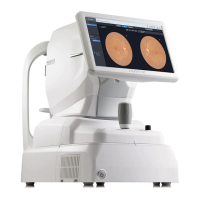
Fundus camera
HFC-1
RE_DATE 2020-06-08
REV R-3 PAGE 32 / 36
Q-I-300-02(R1):2018/06/04 Huvitz Co.,Ltd
11. Compare with other device
Characteristic Fundus camera Fundus camera
Device Name AFC-330 HFC-1
Indication for use
The NIDEK NON-MYDRIATIC AUTO
FUNDUS CAMERA, AFC-330
captures fundus images using a built-
in color CCD camera without the use
of mydriatic agents. The AFC-330 is
useful not only for ophthalmology but
also for fundus photography in
medical examinations such as for
diabetes.
The Huvitz Fundus camera HFC-1 is
a non-contact, bio-microscopic
imaging device. It is indicated for in-
vivo viewing, color fundus imaging.
Intended Use
The NIDEK Non-Mydriatic Auto
Fundus Camera Model AFC-330 is
ophthalmic camera that is indicated
for use in capturing images of the
retina and the anterior segment of the
eye.
A device that illuminates the fundus
by entering light into the pupil and
photographs the fundus state
according to the reflected light.
Observation & photographing of the fundus
Type of photography Color, Red-free& IR Color & Red-free, IR
Angle for view
(33° in Small pupil photography
mode)
45°
Operating distance 45.7 mm 33 mm
Observable/photograph
able diameter of pupil
more
Small pupil diameter : Φ3.3mm or
more
more
Small pupil diameter: Φ3.3mm or
more
Fundus image resolution
Central area :
more
Middle area:
more
Peripheral area: 25 lines pairs/mm or
more
Center :
more
Middle (r/2) :
more
Middle (r) :
more
Observation & photographing of the fundus image
Fixation target
Internal fixation target: Dot matrix
type organic EL
The display position can be
changed and adjusted. The display-
ing method can be changed.
Peripheral fixation target: This is
displayed according to the internal
fixation target displayed position.
External fixation target
Internal fixation target: LCD
External fixation target: White LED
Measurable range of dioptric power for the patient's eye
Without the diopter
compensation lens
Note1
)
-12D to +15D -13D ~ +13D with no compensation
lens
 Loading...