Do you have a question about the Huvitz HFC-1 and is the answer not in the manual?
Indicates the device's intended purpose for in-vivo viewing and color fundus imaging.
Describes the device's function: illuminating the fundus and photographing its state.
Specifies who can operate the equipment, including age, occupation, training, and education.
Details patient requirements for examination, including concentration and following instructions.
Lists conditions or patient types for which the device should not be used.
Provides the Unique Device Identification (UDI) number for the product.
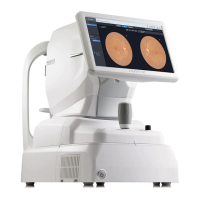
Shows the external appearance of the fundus camera from the front and rear.
Details the names and functions of external components and their corresponding numbers.
Details the names and functions of rear-view components and their corresponding numbers.
Details the names and functions of bottom-view components and their corresponding numbers.
Details the main observation screen layout and the function of each UI element.
Details the confirmation screen for image selection and analysis initiation.
Configures system settings like network, sleep mode, and display.
Configures patient data management, list display, and import settings.
Configures settings for auto tracking, shooting, presets, and image compression.
Adjusts image parameters like brightness, contrast, edge sharpening, and text color.
Configures report logo, brightness, type, pupil size, organization, and image type.
Lists detailed technical specifications of the fundus camera device.
Details essential performance criteria, unacceptable risks, failure modes, and risk controls.
Illustrates the functional connections and components of the device.
Details the function and part code of each component within the device.
Explains critical functional descriptions, features, and operational sequences.
Presents the insulation diagram with creepage and clearance distances for safety.
Outlines the step-by-step workflow for operating the fundus camera.
Diagram illustrating the optical path and components of the fundus camera.
Lists and describes the individual components within the optical system.
Details the light paths for various observation and illumination channels.
Describes the software interface and categories for device calibration and testing.
Details system configuration parameters for device calibration and operation.
Explains factory setup parameters for system LEDs, flash, auto-enhancement, and fixation.
Lists user maintenance tasks such as check-up, cleaning, and replacement.
Provides solutions for common problems encountered with the device's operation.
Instructions for replacing components like chinrest paper and fuses.
Guidance on cleaning the objective lens and illumination lens.
Instructions for cleaning the device exterior and parts contacting the patient.
Covers device storage, cleaning before service, and rubber ring handling.
Details transportation test conditions and requirements for environmental resilience.
| Brand | Huvitz |
|---|---|
| Model | HFC-1 |
| Category | Digital Camera |
| Language | English |