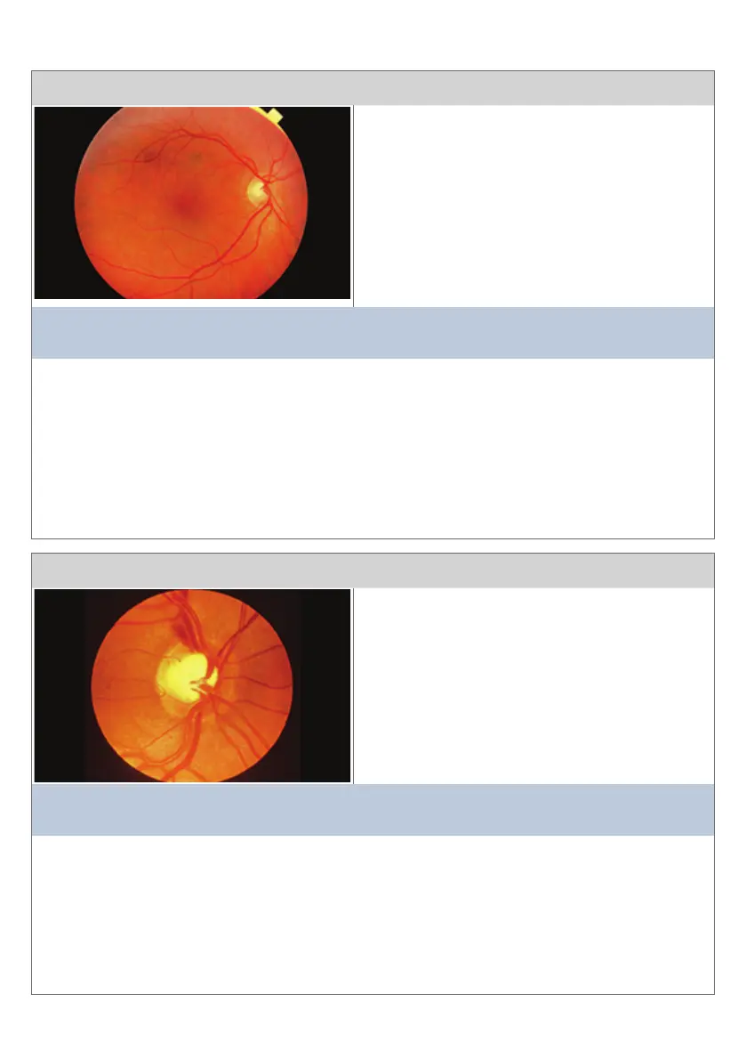
Condion 9
This view is of the opc disc and temporal rena
The main clinical features are:
• Opc disc with uniform central cup with cup
disc rao <0.5 and normal
neurorenal rim
• Renal vessels and macula look normal
• This degree of darker redness in the central
macular area (fovea centralis) is normal
• The slight darkening of the peripheral renal
vessels is also normal
This diagnosis is consistent with:
NORMAL FUNDUS (OPTIC DISC AND RETINA)
Comment:
Sequence for looking at the rena:
• Light reex for cataract, corneal arcus, xanthelesma, conjuncva
• Start at the opc disc
• Superior temporal arcade and inferior temporal arcade
• Macular area
• Superior nasal arcade and inferior nasal arcade
• Peripheral, clockwise sweep to look for peripheral lesions
Condion 10
This view is of the posterior pole centred on the opc disc
The main clinical features are:
• Large cup disc rao (>0.5) indicang cupping
of the opc disc
• Superior polar notching
• Nasal displacement of central blood vessels
This diagnosis is consistent with:
GLAUCOMA
Comment:
Large cup disc rao (>0.5). This means that the cup (central circle) to disc diameter is
> 0.5). The normal rao is 0.3. Tonometry will reveal increased vitreal pressure (usually
greater than 16 mmHg at diagnosis). Glaucomatous damage and its extent is conrmed by
visual elds and tomographic imaging techniques. Central scotoma and a painful eye in most
cases. Consider as cause of severe headache. The diagnosis is an emergency as ongoing
ischaemia due to pressure can precipitate visual loss.
18
Important and Common Renal Condions: Condions 9 - 22
Condions and Diseases of the Rena