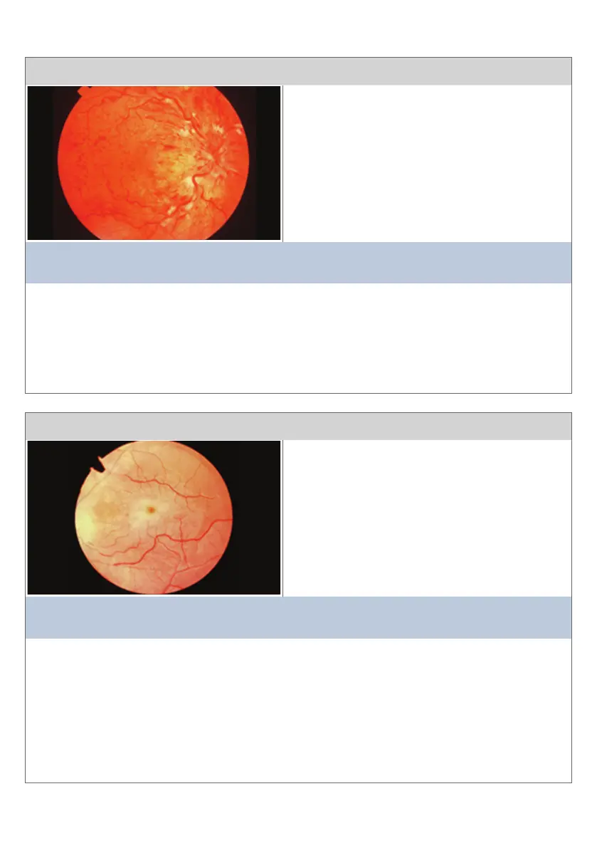
Condion 15
This view is of the opc disc and temporal rena
The main clinical features are:
• Papilloedema
• Tortuosity and dilataon of all branches of
the central renal vein
• Renal haemorrhages: ame shaped, dot
and blot in all quadrants
• Coon wool spots (CWS)
This diagnosis is consistent with:
CENTRAL RETINAL VEIN OCCLUSION (CRVO)
Comment:
The presence of Coon Wool Spots would suggest signicant ischaemic element. Space-
occupying lesions in the cerebrum and hyperviscosity have to be excluded. Hypertension
alone can cause Central Renal Vein Occlusion . The lesions can be beer understood by
considering that arteriolar blood is going into the rena but cannot exit through the central
opc nerve vein.
Condion 16
This view is of temporal rena and temporal opc disc
The main clinical features are:
• Aenuaon of arteries and veins
• The pale temporal edge of the opc disc is
shown
• Central ‘cherry red spot’ with surrounding
pale rena
This diagnosis is consistent with:
CENTRAL RETINAL ARTERY OCCLUSION (CRAO)
Comment:
Poor prognosis due to renal infarcon. Renal cloudiness of pale rena would disappear
aer a few weeks. Aenuated vessels would remain and consecuve opc atrophy would be
evident. The cherry-red spot is seen because the macular arterial supply from the choroid
can remain intact. Oen there is a band of neural ssue that is not rendered ischaemic by
the Central Renal Artery Occlusion, which is seen if there is an adequate cilio-renal artery
supply. This would supply the axons from the fovea centralis renal receptors to the opc
disc.
21


 To Next Page