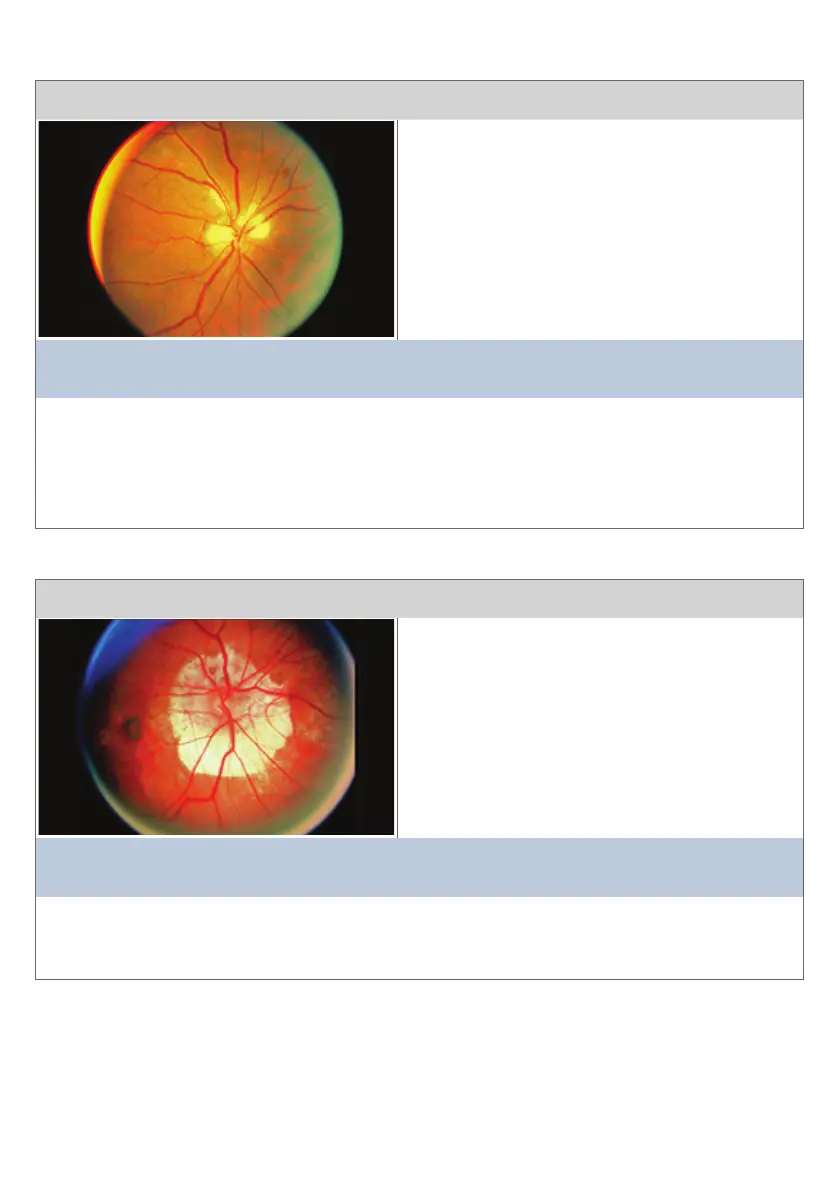
Condion 19
This view is of the opc disc and surrounding rena
The main clinical features are:
• Disc margin and emerging vessels obscured
by myelinated nerve bres along
superior and nasal areas
This diagnosis is consistent with:
MYELINATED NERVE FIBRES
Comment:
On clinical examinaon the blind spot would be expected to be larger, but this would be very
dicult to discern clinically. Approximate 1-3% of the populaon have some degree or other
of this normal variant. During embryogenesis the myelinated nerve bres “spilled” over onto
the renal nerve bres rather that stopping within the opc disc margin.
Condion 20
This view is of the posterior pole centred on the opc disc
The main clinical features are:
• Large opc disc
• Marked peripapillary choriorenal atrophy
This diagnosis is consistent with:
HIGH MYOPIA
Comment:
Areas of choriorenal atrophy in the macular area are not uncommon in highly myopic
paents. The paent would be expected to have an enlarged blindspot.
23