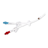
DESCRIPTION
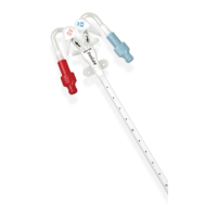
The catheter is divided into two separate round lumens permitting continuous blood ow with one puncture.
All DuoGlide* catheters are made of thermosensitive polyurethane, which softens when exposed to body temperature.
INDICATIONS FOR USE
DuoGlide* Dual Lumen catheters are indicated for use in attaining short term (less than 30 days) vascular access for hemodialysis, hemoperfusion
and apheresis therapy via the jugular, subclavian or femoral vein.
CONTRAINDICATIONS
• Thecatheterisintendedforshort-termvascularaccessonlyandisnottobeusedforanypurposeotherthanindicatedintheseinstructions.
ChloraPrep* Solution One-Step Applicator Contraindications
USA Only
•Donotuseinchildrenlessthan2monthsofagebecauseofthepotentialforexcessiveskinirritationandincreaseddrugabsorption.
•Donotuseonpatientswithknownallergiestochlorhexidinegluconateorisopropylalcohol.
•Donotuseforlumbarpunctureorallowcontactwithmeninges.
•Donotuseonopenskinwoundsorasageneralskincleanser.
WARNINGS
• SUBCLAVIAN ONLY. Pinch-o Prevention: Percutaneous insertion
of the catheter must be made into the axillary-subclavian vein
at the junction of the outer and mid-third of the clavicle lateral
to the thoracic outlet. The catheter should not be inserted into
the subclavian vein medially, because such placement can lead to
compression of the catheter between the rst rib and clavicle and
can lead to damage or fracture and embolization of the catheter.
1
Fluoroscopic or radiographic conrmation of catheter tip
placement should be helpful in demonstrating that the catheter is
not being pinched by the rst rib and clavicle.
1
Signs of Pinch-o
Clinical:
• Dicultywithbloodwithdrawal.
• Resistancetoinfusionofuids.
• Patientpositionchangesrequired
for infusion of uids or blood withdrawal.
Radiologic: (see table)
• Grade1or2distortiononchestX-ray.
Pinch-o should be evaluated for degree of
severitypriortoexplantation.Patients
indicating any degree of catheter distortion
at the clavicle/rst rib area should be followed
diligently.Therearegradesofpinch-othat
should be recognized with appropriate chest
x-rayasfollows:
2,3
• The catheter must not be left in the femoral vein longer than three days. To maintain peak performance it is recommended that
subclavian and jugular catheters be replaced after four weeks.
• Alcohol or alcohol-containing antiseptics (such as chlorhexidine) may be used to clean the catheter/skin site; however, care should be
taken to avoid prolonged or excessive contact of the catheter with the solution(s). Solutions should be allowed to completely dry before
applying occlusive dressing.
• Acetone and Polyethylene Glycol (PEG)-containing ointments can cause failure of this device and should not be used with polyurethane
catheters. Chlorhexidine patches or bacitracin zinc ointments (e.g., Polysporin* ointment) are the preferred alternative.
• Do not resterilize the catheter or components by any method. The manufacturer will not be liable for any damages
caused by re-use of the catheter or accessories.
• Place all clamps near the center of the polyurethane extension pieces. Polyurethane may develop cuts or tears if
subjected to excessive pulling or contact with rough edges. Repeated clamping near or on the Luer-lock connectors
may cause tubing fatigue and possible disconnection.
• Intended for Single Use. DO NOT REUSE. Reuse and/or repackaging may create a risk of patient or user infection, compromise
the structural integrity and/or essential material and design characteristics of the device, which may lead to device failure,
and/or lead to injury, illness or death of the patient.
• Repeated over-tightening of bloodlines, syringes and caps will reduce connector life and may lead to connector failure.
• Enzymes in blood and heparin may cause temporary sticking of the extensions when clamped for extended periods
of time. To release, open clamp and slide away, gently rotating the tubing between ngers and thumb until the tubing separates.
• To avoid damage to vessels and viscus, infusion pressures must not exceed 25 psi (172 kPa). The use of a 10 ml or
larger syringe is recommended because smaller syringes generate more pressure than larger syringes.
NOTE: A three pound (13.3 Newton) force on the plunger of a 3 ml syringe generates pressure in excess of 30 psi
(206 kPa) whereas the same three pound (13.3 Newton) force on the plunger of a 10 ml syringe generates less than
15 psi (103 kPa) of pressure.
• Accessories and components used in conjunction with this catheter must incorporate Luer-lock adapters in order to
avoid inadvertent disconnection.
• Catheters should be implanted carefully to avoid any sharp or acute angles which could compromise the opening of the catheter lumens.
POST DIALYSIS
Use aseptic technique (as outlined above).
1. Flush arterial and venous lumens with a minimum of 10 mL of sterile saline.
WARNING: To avoid damage to vessels and viscus, infusion pressures must not exceed 25 psi (172 kPa). The use of a 10 mL or larger
syringe is recommended because smaller syringes generate more pressure than larger syringes.
2. Inject heparin solution into both the arterial and venous lumens of the catheter. The appropriate heparin solution concentration and
flushing frequency should be based on hospital protocol. Heparin solution of 1,000 to 5,000 units/mL has been found to be effective for
maintaining the patency of hemodialysis and apheresis catheters. When injecting heparin solution, inject quickly and clamp extension
while under positive pressure. Heparin solution volume to lock each lumen must be equal to the priming volume of each lumen. Priming
volumes are marked on each lumen.
3. Clean catheter Luer-lock connectors per hospital protocol. Attach sterile end caps to both the arterial and the venous clamping extension
pieces.
WARNING: To prevent systemic heparinization of the patient, the heparin solution must be aspirated out of both lumens immediately prior
to using the catheter. In most instances, no further heparin solution injection is necessary for 48-72 hours, provided the catheter has not
been aspirated or flushed.
CATHETER REMOVAL
After removing the catheter, apply manual pressure to the puncture site for 10-15 minutes until no signs of bledding are present. Then apply
dressing for 8 hours.
DISPOSAL
After use, this product may be a potential biohazard. Handle and dispose of in accordance with accepted medical practice and all
applicable local, state and federal laws and regulations.
TROUBLESHOOTING
PATIENT WITH FEVER
Patient with fever and chills following the procedure may be indicative of catheter-related bacteremia. If bacteremia is present, removal of the
catheter may be indicated.
INSUFFICIENT FLOW
Excessive force must not be used to flush an obstructed lumen. Insufficient blood flow may be caused by an occluded tip resulting from a clot
or by contacting the wall of the vein. If manipulation of the catheter or reversing arterial and venous lines does not help, then the physician
may attempt to dissolve the clot with a thrombolytic agent (e.g., TPA, Cathflo* Activase* thrombolytic). Physician discretion advised.
CATHETER EXCHANGE
Do not routinely replace dialysis catheters to prevent catheter-related infections
11
. It may become necessary to exchange the indwelling
catheter due to a persistent rise in pressures or decrease of flow rates which cannot be rectified through troubleshooting. Catheter
exchanges should be performed under strict aseptic conditions in which the physician should wear a cap, mask, sterile gown, sterile gloves,
and use a large sterile drape to cover the patient.
REFERENCES
1 Aitken,D.R.andMinton,J.P.“ThePinch-OSign:AWarningofImpendingProblemswithPermanentSubclavianCatheters”,
AmericanJournalofSurgery,Vol.148,Nov.1984,pp.633-638.
2 Hinke,D.H.;Zandt-Stastny,D.A.;Goodman,L.R.;etal.Pinch-osyndrome:Acomplicationofimplantablesubclavianvenous
accessdevices.Radiology177:353-356,1990.
3 Ingle,Rebecca,;Nace,Corinne,VenousAccessDevices:CatheterPinch-oandFracture,1993,Bard Access Systems
4 Sulek,CA.,Blas,ML.,Lobato,EB.,“Arandomizedstudyofleftversusrightinternaljugularveincannulationinadults.”JClin
Anesth.2000Mar;12(2):142-145.
5 Mickley,V.,“Centralvenouscatheters:manyquestions:fewanswers”,NephrolDialTransplant,(2002)17:1368-1373.
6 Tan,P.L.,Gibson,M.,“CentralVenousCatheters:theroleofradiology”,ClinRad.2006,61:13-22.
7 TheInstituteforHealthCareImprovement,“How-to-Guide:PreventCentralLineInfections,”2006.
8 NationalKidneyFoundationK/DOQIGuidelines,2006.
9 TheJointCommissionHospitalAccreditationOrganization,NationalPatientSafetyGoals,2009.
10CenterforDiseaseControlandPrevention,“GuidelinesforthePreventionofIntravascularCatheter-RelatedInfections,”
MorbidityandMortalityWeeklyReport,Aug.9,2002,51(RR-10),1-32.
11TheSocietyforHealthcareEpidemiologyofAmerica,“StrategiestoPreventCentralLine-AssociatedBloodstreamInfectionsinAcuteCare
Hospitals,”InfectionControlandHospitalEpidemiology,Oct.2008,29(S1):S22-S30.
Otherreferencesavailableuponrequest.
Anissuedorrevisiondatefortheseintructionsisincludedfortheuser’sinformation.Intheeventtwoyearshaveelapsedbetweenthisdateandthe
product use, the user can contact Bard Access Systems, Inc. to see if additional product information is available.
Revisiondate:June,2011.
*Bard, Dualator and DuoGlide aretrademarksand/orregisteredtrademarksofC.R.Bard,Inc.
Allothertrademarksarethepropertyoftheirrespectiveowners.
© 2011C.R.Bard,Inc.Allrightsreserved.
Bard Access Systems, Inc.
605North5600West
SaltLakeCity,UT84116U.S.A.
801-522-5000
ClinicalInformationHotline:1-800-443-3385
OrderingInformation:1-800-545-0890
07284011106R
BardAccessSystems
Dual Lumen Catheter
Instructions For Use
Radiologic Signs of Pinch-O
Grade Severity Recommended Action
Grade0 NoDistortion Noaction.
Grade 1
Distortion present
without luminal
narrowing
Chestx-rayshouldbetakentomonitor
progressionofpinch-otograde2
distortion.Shoulderpositioningduring
chestx-raysshouldbenotedasitcan
contribute to changes in distortion
grades.
Grade2 Distortion present with
luminal narrowing
Removalofthecathetershouldbe
considered.
Grade3 Cathetertransectionor
fracture
Promptremovalofthecatheter.
First Rib
Subclavian Vein
Clavicle
Vertebra
Internal Jugular Vein
Superior Vena Cava
Sternum
Pinch-off Area
Infraclavicular Fossa
Axillary Vein