Page 9 of 23 BIOPAC Systems, Inc.
10166_Rev08 WWW.BIOPAC.COM 9.09.2019
Single-Channel ECG Sensor Implant
The single-channel ECG wireless sensor implant contains a small amplifier, sensor and a battery
encapsulated in medical-grade epoxy. Each electrode is 7-strand braided stainless steel with Teflon
insulation. Each strand is 50 μm in diameter. Making the bare electrode 152 μm in diameter (229 μm
diameter insulated). There are two electrode leads on the ECG sensor, and the routing of these leads in the
animal is shown in Figure 11. Spacing for these leads on the sensor is detailed below. The sensor is shipped
deactivated, and is available with a two-month battery life (2 mo) commonly used for mouse studies, or a
six-month battery life (6 mo) commonly used for rat studies. The output gain of the sensor is set at 2000x
during production (±1.0 mV range). A sensor activator must be used to activate sensors on-site. The
technical details of the sensors are as follows:
Footprint: 8 mm x 9 mm (2 mo), 8 mm x 12 mm (6 mo)
Weight: 2.3 g (2 mo), 4.0 g (6 mo)
Volume: 0.76 cm
3
(2 mo), 1.35 cm
3
(6 mo)
System Gain Options: 2000x -- (±1.0 mV range, 1.0 mV in = 2 V out)
Bandwidth: 0.1 – 200 Hz per channel
Input Referred Noise: < 8.0 μV rms at 100 Hz (11 μV rms at 200 Hz)
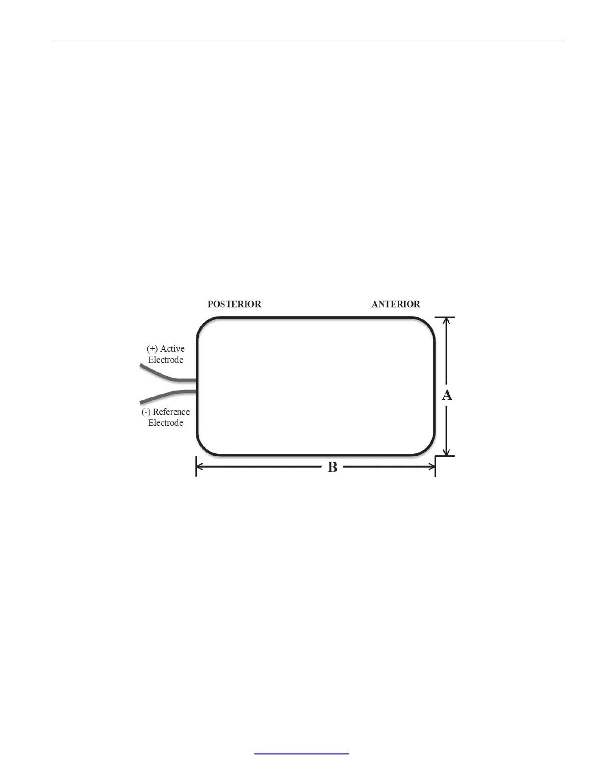
ECG Sensor Electrode Spacing
Figure 11.Single-channel ECG sensor electrode spacing schematic looking down
at animal’s head. The electrodes exit the bottom of the sensor towards the animal’s neck
and back.