Do you have a question about the BK Medical bkSpecto and is the answer not in the manual?
Introduction to the bkSpecto system and how to use this user guide for information.
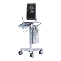
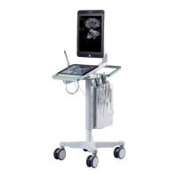
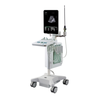
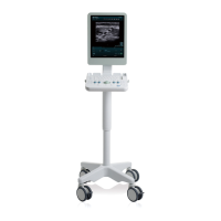
Identifying key system parts and procedures for turning the system on and off.
Steps to start an exam, connect transducers, and manage patient data entry.
Understanding the monitor display, touchscreen controls, and customizing user settings.
Detailed explanation of touchscreen controls for 2D, Color, PW Doppler, and M-Mode imaging.
Parameters and functions for Elastography and 3D imaging, including volume manipulation.
Managing imaging planes, freezing, split screen, and simultaneous imaging features.
Using labels, bodymarks, and arrows to annotate ultrasound images for documentation.
Reviewing recorded cine clips and configuring video output settings for the system.
Step-by-step guide to using caliper tools for measurements in various imaging modes.
Detailed instructions for 2D, Color, and Doppler measurements, including volume calculations.
Overview of document types, saving procedures, and adherence to HIPAA privacy standards.
Managing archived documents, using the browser, and exporting data to external systems.
Process for creating, editing, previewing, and printing patient reports with integrated data.
Optimizing 2D image quality using Focus, Gain, TGC, Zoom, Depth, and Grayscale Map.
Utilizing Tissue Harmonic Imaging, Color/Doppler, Needle Enhancement, and Elastography.
Using Spectral Doppler for flow analysis and M-Mode for motion imaging.
Saving and managing system presets for different imaging applications.
Definition and role of Exam Types in configuring the system for specific examinations.
Performing measurements and Doppler calculations, including RI, PI, and Reduction.
Methods for organ volume calculation and guidance for biopsy and puncture procedures.
Configuring presets, patient data, and imaging controls for prostate exams.
Procedures for biopsy guidance, image annotation, and transducer plane selection.
Steps for measuring prostate volume and generating associated user reports.
Introduction to 3D imaging, modes, license, and transducer movement.
Process for capturing, viewing, and enhancing 3D data sets, including ROI and layouts.
Techniques for rotating, slicing, animating, and annotating 3D volumes for analysis.
Exploring different 3D display modes and performing measurements on 3D data.
Configuring DICOM, managing patient worklists, and understanding DICOM status.
Exporting documents to DICOM printers/PACS and managing DICOM data transfer.
Definitions of key terms and abbreviations used in the user guide and system interface.
An alphabetical list of abbreviations for system measurements and calculations.
Customizing system settings like header, display, patient data, and miscellaneous options.
Managing presets, configuring measurement tools, and assigning labels/bodymarks.
Configuring network, printer, Wi-Fi, and security settings for system operation.
Accessing service details, managing licenses, and viewing system information.
| Type | Ultrasound System |
|---|---|
| Application | Diagnostic Imaging |
| Portability | Portable |
| Probe Frequency | Varies depending on the probe |
| Transducer Frequency Range | Varies depending on the transducer |
| Imaging Modes | B-mode, Color Doppler, Pulsed Wave Doppler |
| Power Supply | AC |
| Connectivity | USB |