Do you have a question about the CEFLA NewTom 5G and is the answer not in the manual?
Provides information about the manual's contents, structure, and conventions.
Describes how the user manual is divided into chapters.
Highlights symbols used for safety information and important notes.
Explains text formatting like bold, italics, and key notations.
Emphasizes following local laws for device installation and maintenance.
Describes symbols found on the device labels.
Explains procedures for system start-up and shut down.
Details the location and use of emergency stop buttons.
Provides essential guidelines for patient and user safety during operation.
Instructions for correctly positioning the patient in the scan area.
Rules for supervising the patient during the scan process.
Procedure for patient exit after exam or emergency shutdown.
How to manually remove a patient if the table malfunctions.
Guidelines on when to repeat a scan due to artifacts or position changes.
Information on risks and safety measures for radiation exposure.
Safety guidelines for laser use during patient positioning.
Requirements for connected peripherals and devices.
Reminder to perform maintenance controls as described in Par. 3.4.
Details on environment, temperature, humidity, and dimensions for installation.
Safety measures against liquid infiltration, fire, ESD, and use of extinguisher.
Rules for modifications and responsibility limits.
Warnings and instructions for device maintenance.
Importance of regular maintenance by authorized personnel.
Procedures and recommendations for cleaning and disinfecting the device.
Guidelines for single-use hygienic protections for patient safety.
Conditions for transport and storage temperature and humidity.
Information for owners and disposal centers on proper equipment disposal.
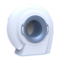
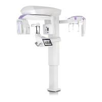
An overview of the NewTom 5G system.
Defines the device's purpose as a cone beam computed tomography x-ray system.
Lists anatomical regions and uses for the system.
Lists prohibited or discouraged uses of the device.
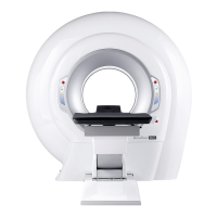
Describes how a patient is positioned and scanned.
Explains the cone-beam technology and scan process.
Details the main components of the system.
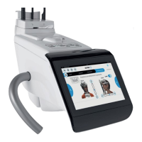
Describes the scanner gantry control panels and indicators.
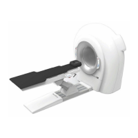
Explains the buttons and functions of the patient table console.
Details the buttons and functions for the patient table with stretcher.
Details the input panel, main switch, and connectors.
Lists standard accessories like QA phantom and calibration support.
Specifies the types of cables included with the device.
Step-by-step instructions for powering on the system.
Step-by-step instructions for powering off the system.
Procedure to prepare the x-ray source, required every two weeks.
Procedure to verify the proper functioning of system parts.
Acquires a background image to calibrate detector performances.
How to invalidate and repeat blank acquisition if an object was present.
Checks proper collimation using a specific test procedure.
Procedures for patient positioning and examination.
Steps to prepare the patient for scanning.
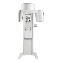
How to position and center a patient for scanning.
Step-by-step guide for positioning a patient on the table.
Guide for positioning patients using the stretcher.
Procedures for scanning a denture.
Steps for positioning a denture for scanning.
Guide for positioning dentures with the patient table.
Guide for positioning dentures with stretcher.
Procedure to properly place and center the QA phantom.
Examples of images obtained from QA phantom analysis.
How to save phantom analysis reports in PDF format.
Refers to a separate NNT – Error Guide document for troubleshooting.
Software utilities and instructions for acceptance testing.
Not applicable for patient support positioning test.
Details laser pointers for patient positioning accuracy.
Refers to manual for section thickness testing.
Instructions for dose measurement according to IEC standards.
Refers to manual for noise and uniformity tests.
Specifies MTF values for spatial resolution.
Technical specifications of the scanner unit, including parameters and dimensions.
Technical specifications of the detector, including pixels, size, depth, and frame rate.
Details image pixel counts and sizes for scout views.
Specifications for reconstructed volumes across different FOVs.
General radiological parameters.
Technical specifications for the X-ray tube model.
Physical and electrical data for the X-ray tube head.
Details on the X-ray source assembly, including cooling curves and rotor specs.
Technical specifications for the inverter unit.
CTDI100 table for operative modalities.
Map showing stray radiation levels.
Technical specifications for the laser used for positioning.
Includes operating temperature, humidity, altitude, and pollution degree.
Guidance and declaration for electromagnetic emissions.
How data is handled during temporary malfunctions.
Lists IEC standards conformity for safety and performance.
Details the plate on the scanner and its position.
Specific label for devices intended for the Chinese market.
Warning label regarding x-ray exposure and unauthorized use.
Warning label for laser device usage.
Warning label for laser devices at specific distances.
Warning label about hand crushing risk.
Label indicating the emergency stop function.
Label providing technical details for the X-ray source.
Label providing technical details for the inverter.
Label stating US federal law restrictions on device sale.
| Type | Cone Beam Computed Tomography (CBCT) |
|---|---|
| Imaging Technology | 3D Imaging |
| Detector Type | Flat Panel Detector |
| Applications | Dental, Maxillofacial, ENT |
| Voxel Size | 0.075 mm |
| Scan Time | 3.6 seconds |
| Radiation Dose | Low dose |
| X-ray Generator | High-frequency generator |
| Software | NNT Software |
| Power Requirements | 220-240 V, 50/60 Hz |