1
Fiber Photometry Systems at a Glance
1.1 Systems Overview
In neuroscience, Fiber photometry denotes a method whereby chronically implanted optical fibers deliver excitation
light to neurons tagged with a fluorescent calcium indicator and collect their overall activity-induced fluorescence. Fiber
photometry sums up the activity-induced fluorescence of all neurons expressing the indicator(s).
In addition to systems custom-designed for your needs, we offer systems for typical modular fiber photometry mea-
surement setups. The Locked-in or Sequential Detection for GCaMP Isosbestic and Functional Excitations system measures
the 405 nm (isosbestic point) excited GCaMP fluorescence, and the 465 nm excited calcium-dependent GCaMP fluo-
rescence, on a single photodetector. The Separated Two Fluorophores Fluorescence contains all the items necessary to
perform photometry measurements of two independent colors in freely-moving animals for GFP-like and RFP-like fluo-
rophores.
A typical set-up for freely behaving animals contains of the following elements (Fig. 1.1).
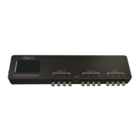
• A Fiber photometry console to synchronize output control and the acquisition of data.
• An Integrated fluorescence mini cube where beam splitters combine the excitation wavelengths and separate the
emission wavelengths. This Fluorescence Mini Cube comes with the following.
– One or more Built-in fluorescence detector heads capable of sensing low-intensity light. The fluorescence emis-
sion can be collected with one photodetector and subsequently demodulated or, after spectral separation,
collected with respective photodetectors.
– One or more Built-in LED optical heads that provide light for the experiment.
• An optional Fiber-optic rotary joint can be used to allow free movement of the experimental subject.
• An Optical cannula, with connecting Fiber-optic patch cords, to deliver light to the subject.
• A Low Autofluorescence Patch Cord to allow light collection with minimal fluorescence noise.
The following section describes these standard elements and their purpose.
Chapter 1. Fiber Photometry Systems at a Glance 3


 To Next Page