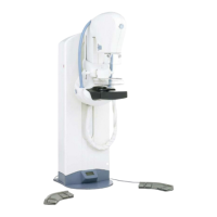
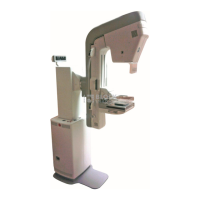
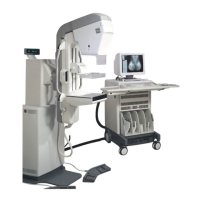
The Senographe 2000 D Acquisition System is a digital mammography system designed for screening and diagnostic breast examinations, including spot compression, magnified, and coned views. It is a modular system that eliminates the need for film cassettes by leveraging digital technology for on-screen image display, networking, filming, and archiving.
Function Description
The system is equipped with a dual-track X-ray tube (molybdenum/rhodium) and a flat panel digital detector of amorphous silicon with cesium iodide, optimized for X-ray detection and light photon transmission. The Control Console manages positioning operations and X-ray exposure, as well as power to all system components. The Acquisition Workstation (AWS) handles image acquisition, processing, and display, database management, and can send images for archiving, review, or filming. The system supports examinations for standing, sitting, or recumbent patients, offering both contact and magnification views.
Important Technical Specifications
The Senographe 2000 D operates continuously with intermittent loading.
- Electrical Specification:
- Single-phase input voltage: 200/208/220/240/380/415 V (± 10%).
- Can be powered by single-phase 440 V (± 10%) using an optional transformer.
- Line frequency: 50 or 60 Hz (± 5%).
- Protection against electrical shocks: Class 1, Type B.
- Maximum line current corresponds to 30 kV, Mo track, large focal spot, and 100 mAs.
- kVA Load Characteristics:
- Maximum power in standby: 1.5 kVA.
- Maximum instantaneous power (during exposures, up to 6 seconds): 9 kVA.
- Power factor: 0.6.
- Line current crest factor: 1.7 at 200 V to 2 at 415 V.
- Input impedance: The apparent resistance of the mains supply (RL) must be less than that which would cause a voltage drop of 6% at the maximum power load of 9 kVA. For 380 V, RL(380) < 1 Ω. For other voltages (U), RL(U) < (U/380) Ω.
- Generator Output (excluding tube):
- 22 through 49 kV.
- 20 through 130 mA.
- Duty Cycle: The generator's duty cycle is always limited by the X-ray tube.
- Maximum Tolerance of Displayed Constants:
- kV: ±5%.
- mAs: ±7.6% ±1.1 mAs.
- Digital Detector FOV: 19 cm x 23 cm.
- Radiation and Filter Information:
- Radiation reference axis: Directed at the chest wall edge of the digital detector, with radiation shielded behind the chest wall.
- Technical leakage factor: Compliant with DHHS 21 CFR1020 (49 kV at 2 mA).
- Filters and Anode Tracks: Molybdenum (Mo) and Rhodium (Rh) tracks.
- Large focal spot: 100 mA max (Mo), 75 mA max (Rh).
- Small focal spot: 40 mA max (Mo), 40 mA max (Rh).
- Filters: Molybdenum (0.03 mm) and Rhodium (0.025 mm).
- Half-Value Layer (HVL) at 30 kV: 0.03 Mo filter (0.3 mm Al minimum), 0.025 Rh filter (0.35 mm Al minimum for Mo track, 0.4 mm Al minimum for Rh track).
- Minimum permanent filtration: 0.008 mm Al (8 μm Al) at 30 kV (0.69 mm Beryllium).
- Ambient Conditions:
- Operational: Humidity 10-80% max, Temperature 15-35°C (59-95°F) max, Atmospheric pressure 700-1060 hPa max.
- Transport and Storage: Humidity 10-50% max (95% max for two weeks), Temperature +10-25°C (50-77°F) max (-10-50°C (14-122°F) for one day), Atmospheric pressure 500-1060 hPa max.
- Ambient Light Level: Monitors adjusted for optimum 50 lux.
- Workstation Storage: Total internal disk capacity: two 9 Gbyte disks. Allocated for image storage: 14 Gbytes (approx. 1650 images).
Usage Features
- AOP (Automatic Optimization of Parameters) Mode: This mode automatically selects optimal radiographic parameters (track, filter, kV, and mAs) based on breast thickness and density, prioritizing contrast, dose reduction, or a standard compromise. Breast compression of at least 3 daN (30 Newtons or 6.7 pounds) is essential for AOP mode. Markers larger than 2 mm² should not be placed in the AOP algorithm's area to avoid affecting tissue density calculations. AOP mode is not recommended for patients with mammary implants.
- Manual Mode: Allows operators to manually select focal track (Mo/Rh), filter (Mo/Rh), kV, and mAs values.
- Compression System: Features a magnetic braking mechanism to prevent paddle fall in case of power loss. If power loss occurs during compression, a force of around 5 daN remains, requiring gentle manual release. Upward movement of the compression paddle stops if a downward force greater than 3 daN is applied, minimizing injury risk.
- Image Receptor: The digital detector is integrated into the Image Receptor, which serves as a breast support. It includes a removable grid (Bucky) that can be interchanged with an optional breast holder without a grid. Magnification platforms are available, automatically selecting the small focal spot.
- Gantry Control Console: Provides operator commands, displays system replies and messages, and controls power. It includes switches for system power, gantry power off, SETUP/MEDICAL menu access, readout display, focal spot/track selection, filter selection, breast laterality selection, special views, kV/mAs selection, exposure prep, exposure, and compression release.
- Acquisition Workstation (AWS) Cart: Houses the AWS monitor, keyboard, mouse, computer, electronics, accessory storage, and an Uninterruptible Power Supply (UPS). The three-section table (keyboard, light box, writing surface/mouse pad) can be raised for access to accessories and its sides can be lowered.
- Bar Code Scanner (Optional): A fast and accurate tool for scanning bar code symbols (Code 3 of 9, EAN 13, Code 128) to automatically enter patient information into the AWS, reducing manual entry errors.
- Image Acquisition Procedure: Involves patient/exam selection from the Worklist, equipment preparation (image receptor, field size, compression paddle, exposure mode), patient positioning (removing folds, positioning nipple, bringing out inframammary angle, compression), and image acquisition.
- Image Processing: Raw images are acquired and immediately displayed, then processed to create high-quality images. Processing includes collimator detection (black mask around useful area), pseudo-log transformation (dynamic range reduction to 12 bits), and either Thickness Equalization or Premium View (optional). Thickness Equalization enhances visibility in all breast regions, while Premium View optimizes visibility of breast structures.
- Image Review: Acquired images are stored on an internal disk for preliminary assessment and can be reviewed on the AWS monitor. The Viewer window allows display, manipulation (zoom, 2D localization, contrast/brightness adjustment, image inversion, geometric transformations), and annotation of images.
- Printing: Supports printing of processed images to GE-recommended high-resolution DICOM laser cameras. Auto Print automatically prints images with an "OK" Quality Check value upon exam closure. Manual Print allows printing individual images from the Viewer. Only films formatted for a single mammogram per sheet are recommended for diagnostic purposes, and only 8x10 inch films have been validated.
- Interchange Media (Optional CD-R Drive): Allows saving and restoring Senographe 2000 D image data on standard 5-1/4” recordable compact disks (DICOM format) for communication purposes. Not recommended for permanent archiving.
Maintenance Features
- Quality Control: Regular QC procedures are recommended, as described in the Senographe 2000 D QAP QC Manual, to ensure continued high-level operation.
- Planned Maintenance: Performed by a Radiologic Technologist (daily to semi-annually) for simple QC tests, by a Medical Physicist (annually) for mammographic quality, and by a GE Service Representative (twice a year) for comprehensive checks.
- Cleaning and Disinfection: Instructions are provided for cleaning the monitor screen and housing using a microfiber cloth with water or ethyl alcohol. Patient contact surfaces (bucky/Image Receptor, compression paddles, magnification platform, face shield) require low or intermediate level disinfection using suitable liquid chemical germicides. High-level disinfection is recommended for contact with broken skin or infected patients. Specific germicides like VESPHENE IIse and CIDEX are recommended, with strict adherence to manufacturer instructions.
- Error Messages: The system displays error messages on the Column Readout, Control Console, or AWS screen to indicate operational anomalies. These messages provide explanations and recommended corrective actions, ranging from simple resets to contacting GEMS Field Service.
- UPS (Uninterruptible Power Supply): Integrated into the AWS cart to ensure continuous power during fluctuations or brief interruptions, allowing for orderly shutdown and data protection in case of power failure. Regular checks of UPS function and battery capacity are part of planned maintenance.