Do you have a question about the Hologic Fluoroscan InSight FD Mini C-arm and is the answer not in the manual?
Explains the nature of X-ray emission and primary/secondary radiation.
Details radiation dose, dose rate, and factors affecting exposure.
Details compliance with US CFR and state-specific radiation regulations.
Specifies compliance with relevant International Electrotechnical Commission (IEC) standards.
Details the location and content of the main system label.
Explains the X-ray system warning label and its importance.
Explains how the Field of View is indicated and selected.
Details risks associated with radiation and actions to mitigate them.
Highlights electrical hazards and safety measures for high voltages.
Explains the risk of the system tipping over and how to prevent it.
Explains potential causes for imaging function loss.
Warns against unauthorized modifications to the X-ray tube.
Provides guidance on actions to take in case of an electrical fire.
Outlines precautions to take in potentially explosive atmospheres.
Addresses potential hazards related to the foot switch.
Explains collision risks during movement and how to prevent them.
Details safe cleaning practices and recommended cleaning agents.
Warns about connecting unapproved devices to the system.
Discusses potential effects of electrosurgical devices on system operation.
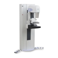
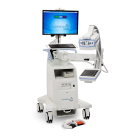
Labels and describes the key physical parts of the InSight FD system.
Describes the functions of buttons and symbols on the X-ray head panel.
Lists keyboard shortcuts and their corresponding functions for system operation.
Step-by-step instructions for safely moving the InSight FD system.
Instructions on how to adjust the C-arm's height and position.
Guidance on fitting a sterile drape to the C-arm for sterile procedures.
Steps for powering on the system and its conditioning schedule.
Procedure for safely shutting down the InSight FD application and system.
Describes the multi-page tab-selected window for system settings.
Details the main system settings accessible via the configuration menu.
Explains the fields and options available on the System page.
Configures destinations for sending DICOM images and data.
Sets up destinations and parameters for printing DICOM images.
Configures the system to access and query worklists from providers.
Details how to add, edit, and manage user accounts for system access.
Allows administrators to set default preferences for physicians.
Configures default image acquisition settings for physicians.
Configures default image processing settings for physicians.
Configures default image management and printing preferences.
Manages system logs, audit mode, touch screen, and shutdown features.
Describes the main screen layout and available menus/buttons.
Details the process of acquiring new X-ray images.
Guides users on selecting or entering patient information.
Explains the layout and controls of the image acquisition screen.
Process for reviewing previously acquired images.
Guides users in selecting studies for review from the system.
Interface for viewing and managing acquired images.
Provides detailed viewing options for individual images.
Managing patient and study information within the system.
Procedure for updating patient and study data.
Instructions for exporting images from the system.
Steps to export images directly from the Review Images screen.
Procedure for exporting images using the Tools Menu.
Guides users on importing images into the system.
Procedure for selecting and deleting images from the system.
Displays the status of DICOM send requests and allows retries.
How to back up system configuration and calibration data.
How to restore system configuration and calibration data.
Lists approved cleaners and disinfectants for the system.
| Type | Mini C-arm |
|---|---|
| Image Acquisition | Digital |
| X-Ray Tube Type | Stationary Anode |
| Frame Rate | 30 fps |
| Monitor | High-Resolution LCD |
| Power Requirements | 100-240 VAC, 50/60 Hz |
| Applications | Orthopedic |
| Detector Type | Flat Panel Detector |
| Monitor Size | 24" (61 cm) |
| Field of View | 8" x 10" (20 cm x 25 cm) |
| X-ray Tube Voltage | 40 - 75 kVp |
| Image Processing | Real-time image processing |