C-Arm Subsystem
Circuit/Mechanical Descriptions
The following paragraphs describe basic electrical and physical features of the 9800 SERIES C-Arm Subsystem (referred to
hereafter as the C-Arm). Subsequent chapters in this manual explain subsystem operation in functional concepts, focusing on
how subsystem components interact in providing those functions. You can obtain additional information from the C-Arm
Interconnect Diagram and Operators Manual, and the Workstation Operators and Service Manuals.
Subsystem Description
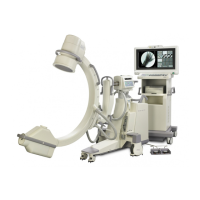
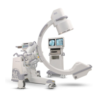
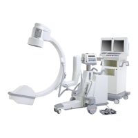
The C-Arms derive their name from the rigid, C-shaped weldment that supports the Image Intensifier, collimator, CCD Camera,
and X-ray tube. These components together are commonly called the “C”, and the entire assembly from the casters up is known
as the “C-Arm”.
The X-Ray tube generates an X-ray beam that is shaped and oriented by the collimator. After passing through patient anatomy,
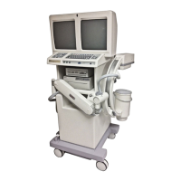
the beam is directed onto a film cassette to produce a radiographic film image, or through the image intensifier and CCD camera
(charge-coupled device), to convert the X-ray image to a video signal that is transmitted to the Workstation for display,
processing, and storage.
There are several options and variations of C-Arm (see table following). Image Intensifiers are available in 9-inch and 12-inch
sizes. The larger, more expensive II is often necessary for cardiac and vascular studies that require images of larger areas of the
body. The smaller II is useful in extremity imaging and other routine applications.
The Super C is often used in cardiac studies that require oblique views through the body, although it has proven useful in other
applications as well. The Super C-Arm is similar to the standard C-Arm except that it has a larger diameter C, which mounts
directly on the end of the Cross Arm. Due to geometric characteristics, there are variations in the types of motions of which each
is capable (see following table).
A Cardiac C-Arm must be able to generate high technique in a pulsed mode to support dynamic cardiac and vascular studies.
These studies can be recorded to disk or tape and replayed showing detailed, real-time movement. The Cardiac C-Arm capacity
is increased by software options that enable the generator to produce higher technique X-ray pulses than the standard C-Arm.
2
Service
Periodic Maintenance
Contents
Schematics
Illustrated Parts
Installation
 Loading...