Condion 4
This view is of the central fundus with the opc disc
The main clinical features are:
• New vessels on the disc (NVD) and else-
where (NVE) along the vascular arcades
• Haemorrhages
• Hard exudates
• Pre-renal brosis
This diagnosis is consistent with:
ADVANCED PROLIFERATIVE DIABETIC RETINOPATHY
Comment:
On-going ischaemia and increase in vaso-proliferave factors. The New Vessels grow into
the vitreous and are fragile leading to haemorrhage. As the haemorrhage organises, brous
ssue reacon occurs oen resulng in traconal renal detachment.
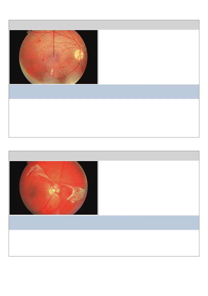
Condion 3
This view is of the opc disc and temporal rena
The main clinical features are:
• Mulple dot and blot haemorrhages
• Coon wool spots (CWS)
• Intra-renal micro-vascular abnormalies
(IRMA)
• New Vessel formaon on the disc (NVD)
This diagnosis is consistent with:
PRE-PROLIFERATIVE DIABETIC RETINOPATHY
Comment:
Pre-proliferave Diabec Renopathy is characterised by renal ischaemia. Coon
Wool Spots represent areas of focal renal ischaemia and Intra-renal micro-vascular
abnormalies are a pathological aempt at micro-revascularisaon. Intra-renal micro-
vascular abnormalies are at and do not grow into the vitreous. The tangle of ne blood
vessels at the opc disc may be early New Vessels on the disc.
15