Condion 6
This view is of the central fundus with the opc disc
The main clinical features are:
• Focal areas of pigmentaon consistent with
focal laser photocoagulaon
• Mulple hard exudates are seen within the
macular area
• Some in a circinate paern with central
Microaneurysms
This diagnosis is consistent with:
DIABETIC MACULOPATHY: ONGOING WITH PREVIOUS FOCAL LASER PHOTOCOAGULATION
Comment:
Visual acuity will almost certainly be ≤ 6/12. Microaneurysms leak plasma which precipitates
in a lipid-rich protein deposit which is hard exudate. The circinate nature of some hard
exudates with central Microaneurysms clearly suggests this. The central Microaneurysms
within a clump or circinate of exudates are laser photocoagulated to obliterate and seal the
Microaneurysms to reduce plasma leakage.
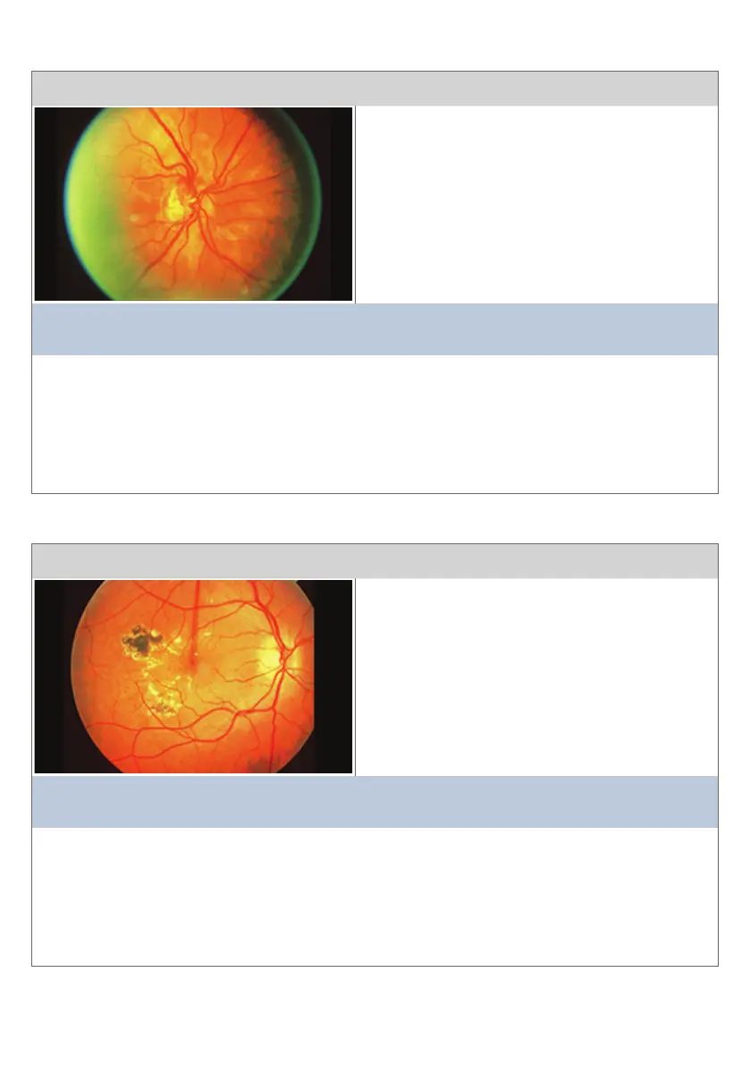
Condion 5
This view is of the opc disc
The main clinical features are:
• New Vessels formaon on the disc (NVD)
• Myopic Crescent over the temporal edge of
the opc disc
• Peripheral renal pigment layer prominence
This diagnosis is consistent with:
PROLIFERATIVE RETINOPATHY WITH NEW VESSELS ON THE DISC (NVD)
Comment:
New Vessels on the disc are oen dicult to see and the rst sign of Proliferave Diabec
Renopathy. Myopic Crescent over the temporal edge of the opc disc is subtle with
peripheral renal pigment layer prominence (oen seen in Myopia). This should not be
confused with Renis pigmentosa. The New Vessels will grow further into the vitreous and
are fragile and can lead to haemorrhage.
16
Condions and Diseases of the Rena (Connued)