Do you have a question about the Chison ECO 3 EXPERT VET and is the answer not in the manual?
Discusses measures to ensure operator and patient safety, including general user precautions.
Details protection against electric shock, water ingress, and safety levels for flammable atmospheres.
Discusses safe usage of diagnostic ultrasound equipment and principles for minimizing biological effects.
Outlines principles for using ultrasound for medical diagnosis by trained personnel.
Explains MI and TI output displays for assessing thermal and cavitation bioeffects.
Details the steps and requirements for installing the ultrasound system.
Guides on acclimation time, connecting electric power, and system boot procedures.
Provides instructions and cautions for correctly installing ultrasound probes.
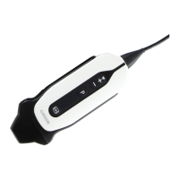
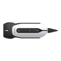
Explains the purpose and operation of various function keys on the control panel.
Details the central control elements and their multifunction operations with the trackball.
Describes controls for adjusting image parameters like THI, AIO, and CINE functions.
Outlines steps for device inspection, checking voltage, connections, and probe status.
Details the process of entering patient information, including function buttons and operation.
Explains adjustments for B mode images like Frequency, Dynamic range, and i-Image.
Guides on increasing or decreasing image frequency.
Explains adjusting contrast resolution by changing dynamic range.
Details the i-Image function for image adjustment.
Explains the Compound imaging function for adjustment.
Details the SRA function for turning it on or off.
Guides on adjusting M speed in real-time M status.
Guides on adjusting B Image Menu parameters like Scan Width, Focus Num, Persistence.
Explains adjustments for CFM mode image parameters like Color Map and Contrast.
Details adjustments for PW mode image parameters like Enhance and D Gamma.
Provides instructions and cautions for cleaning the ultrasound machine.
Details care, maintenance, cleaning, and disinfection procedures for ultrasound probes.
Describes regular checks to ensure the device works normally and safely.
Lists common malfunctions, their reasons, and corresponding measures for troubleshooting.
Outlines daily inspection, handling, cleaning, and disinfection precautions for probes.
Details how to inspect probes for damage before and after each use.
Provides procedures for cleaning and disinfecting various types of ultrasound probes.
Provides detailed cleaning and disinfecting procedures for transvaginal and transrectal probes.
| Probe Ports | 2 |
|---|---|
| Ports | USB, VGA, HDMI |
| Type | Ultrasound System |
| Image Modes | B, M |
| Transducer Frequency | 2.5-10 MHz |
| Storage Capacity | 500GB HDD |
| Applications | Veterinary use |
| Probe Type | Convex, Linear, Micro-convex |
| Scan Modes | Color Doppler |
| Measurement Functions | Distance, Area, Volume, Heart Rate |
| Power Supply | 100-240V~ 50/60Hz |