
M
Melissa MartinezAug 5, 2025
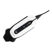
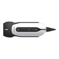
What to do if the menu bar displays but there is no scanning image on Chison SonoEye P2 Medical Equipment?
- BBenjamin SmithAug 5, 2025
If the menu bar is visible but there's no scanning image on your Chison Medical Equipment, it may be due to incorrect transmission frequency, gain, or STC control settings. Adjust these settings accordingly. Also, ensure that the system is correctly connected. If the system is frozen, defreeze it by pressing the FREEZE icon.