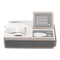
SE-3 Electrocardiograph Service Manual Operating Principle
- 13 -
Chapter 3 Operating Principle
This chapter describes the basic theory and the internal circuit structure of SE-3 to let the service
provider understand the operating principle.
3.1 Basic Theory of ECG Operation
The heart is a power organ of the blood circulation. Before the systole or the diastole, a cardiac
impulse happens in the heart muscle, and a faint bioelectric signal is thus generated. The
bioelectric signal is transmitted through the whole body, and the potential difference is generated
on the different skin surfaces because of the different distances from the heart.
The cardiogram is a record of the amplification of the potential distribution on the body skin
surface. The potential difference is sampled by the electrodes, and amplified and processed by the
electrocardiograph. Then it is recorded on the paper. The cardiogram recorded by the
electrocardiograph can help doctors to analyze and diagnose heart disease. The intended use of
the electrocardiograph is to acquire ECG signals from adult and pediatric patients through body
surface ECG electrodes. The electrocardiograph only records the heart's electrical activity, and
does not produce any electricity of its own. The test does not hurt and has no known side effects.
It does not require any preparations except possibly shaving chest hair to get a better recording.
The recording itself takes only a few seconds.
It is more than one hundred years since the electrocardiograph was applied in clinical diagnosis.
The electrocardiograph is an important measurement in clinical diagnosis of heart disease, and is
equipped in almost every hospital and clinic.
3.2 Composition of ECG
The standard 12-lead electrocardiogram is a representation of the heart's electrical activity
recorded from electrodes on the body surface. A normal ECG wave consists of a P wave, a QRS
complex, an ST segment, a T wave and a U wave. In the following figure, the x-axis indicates
time and y-axis indicates voltage. When the recording speed is 25mm/s and the sensitivity is
10mm/mV, one small grid on x-axis represents 0.04 seconds, and one small grid on y-axis
represents 0.1mV.
This diagram illustrates ECG waves and intervals as well as standard time and voltage measures
on the ECG paper.