CHAP. 1
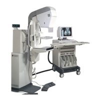
GE Healthcare Senographe 2000 D Acquisition System
REV 1 OM 5179217–1–100
18
8. RESIDUAL IMAGES
Repeated exposures made with a high contrast object in the digital detector area may lead to the
creation of a ghost image, caused by an excessive difference of residual charges between cells of the
detector. The time taken for this ghost image to disappear depends upon the magnitude of the
residual charges.
This effect can occur during checks to measure the X-ray field which use a dosimeter ionization
chamber in the beam. To avoid it, use one of the following solutions:
D Reduce the contrast of the object by including it in a field protected by an X-ray attenuator of
adequate thickness.
D Use the internal dose measurement facility.
D Protect the digital detector by means of a 3 mm steel plate for the duration of the measurements.
9. DAMAGE TO THE DIGITAL DETECTOR
The digital detector contains thallium doped cesium iodide, a substance which requires special
precautions for handling and recycling. If the protective casing of the digital detector sustains damage,
please consult your local GEMS representative.
If the digital detector casing is punctured, the detector must be removed by
authorized GE Service personnel wearing protective gloves and dust masks;
send the protective items for disposal along with the defective detector.
CAUTION
FOR TRAINING PURPOSES ONLY!
NOTE: Once downloaded, this document is UNCONTROLLED, and therefore may not be the latest revision. Always confirm revision status against a validated source (ie CDL).
 Loading...