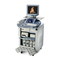
GE HEALTHCARE - KRETZTECHNIK VOLUSON® 730EXPERT (BT03)
D
IRECTION 105899, REVISION 3 DRAFT (APRIL 29, 2008) SERVICE MANUAL
Chapter 5 - Components and Functions (Theory) 5-17
5-2-5-4 VOCAL - Virtual Organ Computer-aided Analysis
Diagnosis and therapy of cancer is one of the most important issues in medical care.
The VOCAL™- Imaging program is an extension of the 3DView™ software, integrated in the Voluson®
sonography systems and also available for PC. It allows completely new possibilities in cancer
diagnosis, therapy planning and follow-up therapy control.
VOCAL™ offers different functions:
Volume Calculation - Manual tracing of contours in three dimensions
3D Color Histogram - Automatic calculation of the vascularization
Shell Imaging - construction of a virtual shell which covers the entire contour to separately calculate
internal tumor vascularization and peripheral vascularization for tumor therapy planning and follow up
control.
5-2-5-5 B-Flow
B-Flow is especially intuitive when viewing blood flow, for acute thrombosis, parenchymal flow and jets.
It helps to visualize complex hemodynamics and highlights moving blood in tissue.
B-Flow is less angle-independent, no velocity aliasing artifacts, displays a full field of view and provides
better resolution when compared with Color-Doppler Mode. It is therefore a more realistic (intuitive)
representation of flow information, allowing to view both high and low velocity flow at the same time.
5-2-5-6 XTD-View (Extended View)
XTD-View provides the ability to construct and view a static 2D image which is wider than the field of
view of a given transducer. This feature allows viewing and measurement of anatomy that is larger than
what would fit in a single image. XTD-View constructs the extended image from individual image frames
as the operator slides the transducer along the surface of the skin in direction of the scan plane.
Examples include scanning of vascular structures and connective tissues in the arms and legs.
5-2-5-7 DiagnoSTIC (Spatio-Temporal Image Correlation)
With this acquisition method the fetal heart or an artery can be visualized in 4D.
It is not a Real Time 4D technique, but a post processed 3D acquisition.
• DiagnoSTIC - Fetal Cardio is only available on RAB & RIC probes in the OB/GYN application.
• DiagnoSTIC - Vascular is only available on the RSP probe in the Peripheral Vascular application.
5-2-5-8 CRI - Compound Resolution Imaging
In this special B-mode, beams are transmitted not only perpendicularly to the acoustic window, but also
in oblique directions. Between three and nine beams are correlated to form one image line.
The advantages of Compound Resolution Imaging are enhanced contrast resolution with better tissue
differentiation and clear organ borders. Also vessel walls and tissue layers are emphasized for easier
recognition.
5-2-5-9 VCI - Volume Contrast Imaging
Volume Contrast Imaging utilizes 4D transducers to automatically scan multiple adjacent slices and
delivers a real-time display of the ROI.
This image results from a special rendering mode consisting of texture and transparency information.
VCI improves the contrast resolution and therefore facilitates finding of diffuse lesions in organs.
VCI has more information (from multiple slices) and is of advantage in gaining contrast due to improved
signal/noise ratio.

 Loading...