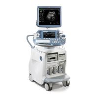
Do you have a question about the GE Voluson Swift and is the answer not in the manual?
Provides contact information and support resources for the system.
Information about the developer and producer of the Voluson™ SWIFT / Voluson SWIFT+ system.
Details on standards and regulations the Voluson™ SWIFT / Voluson SWIFT+ conforms to.
Guidance on how to use and understand the Instructions for Use document.
Information on the system's intended use, clinical applications, patient population, and operator profile.
Description of all symbols and labels used on the system and in the Instructions for Use.
Key precautions and warnings for safe operation of the ultrasound system.
Safety guidelines and requirements for electrical installation of the system.
Precautions regarding environmental conditions for safe operation and potential interference.
Instructions and cautions for safely moving the ultrasound system, including lifting.
General safety precautions to be observed during system operation.
Instructions for cleaning the ultrasound device, including approved agents and methods.
Information on recommended maintenance schedules and safety tests for the system.
Guidelines for the proper disposal of the system and its components, including batteries.
Information on the risks of ultrasound waves, ALARA principle, and bioeffects.
Declarations related to electromagnetic emissions and immunity according to standards.
Specifications and requirements for network connections and protocols used by the system.
Information on proactive measures taken to secure the system and recommendations for users.
Details on how GE field engineers access the system remotely for diagnosis and maintenance.
Procedure for downloading and installing software updates for the system.
Information on messages related to high system temperature and potential shutdown.
General description of the Voluson™ SWIFT / SWIFT+ as a professional ultrasound scanning system.
Diagram and list of the main components and parts of the ultrasound system.
Details on the system's user interface, including console adjustments and controls.
Description of the control panel layout and the function of each button.
Information on monitor adjustment, display layout, and control areas.
Step-by-step instructions for powering the system on, off, and using sleep mode.
Instructions on connecting and selecting a probe, entering patient data, and changing presets.
Explanation of fundamental operations like Gain, Zoom, Depth, Focus, and Automatic optimization.
General safety precautions and warnings for handling and using ultrasound probes.
Recommendations and procedures for cleaning and disinfecting ultrasound probes.
Information on probe intended use, contraindications, patient population, and labeling.
Safety precautions and procedures related to biopsy setup, mounting, and use.
Details on specific 3D/4D and 2D probes, including clinical applications and features.
Instructions on how to use the probe assessment tool and understand its acceptance criteria.
Description of the components of the 2D Mode screen display, including markers and scales.
Explanation of standard features and modes available in 2D Mode, such as B-Mode, CFM, and PD.
Details on available 2D Mode options including CW Doppler, Extended View, and Contrast Media.
Explanation of how to adjust the TGC slider controls to vary gain in the 2D image.
Description of the Scan Assistant tool, its sections, and how to activate and edit it.
Instructions on how to add text and indicators to ultrasound images, including editing and exiting modes.
Information on how to use Cine Mode for reviewing image sequences, including editing and split functions.
Explanation of the principle of volume acquisition and the standard representation of sectional planes.
Tips for achieving optimal 3D/4D image quality, focusing on B-Mode and ROI placement.
Explanation of resetting rotations and translations of a volume section to the initial position.
Description of the components of the 3D/4D Mode screen display, including Volume Box and Render Box.
Information about the Volume Pre mode, including sub menus and controls for acquisition.
Details on 4D Real Time, 3D Static, and VCI-A acquisition modes for volume data.
Overview of visualization modes like Render, Multiplanar, TUI, OmniView, and their controls.
Description of additional tools such as Render Modes, MagiCut, and Cine Calculation for image manipulation.
Instructions on how to open the Archive to view, print, and transfer images and data.
Overview of data transfer controls, including export, import, and sending data via DICOM or Email.
Procedures for uploading data to Tricefy and sending data via email.
Details on exporting images and volumes in various formats to external devices or networks.
Functionality to anonymize patient, exam, and image data for privacy protection.
Instructions for importing data from USB devices or DICOM servers into the local archive.
Procedure for deleting patients, exams, and images from the archive, including warnings for locked items.
Details on setting up and using the DICOM server for data transfer and retrieval.
Controls for managing patient information, including input fields and data entry.
Instructions for using the Worklist to search for and manage patient exams.
Dialog for entering data from previous ultrasound exams performed on other systems.
Description of the clipboard's functionality for displaying and managing preview images.
Functionality to compare current images with images from previous exams.
Overview of the measurement menu, including groups, available measurements, and study navigation.
Details on basic operations and available generic measurements like distance, area, angle, and vessel measurements.
Information on how to perform calculations based on measurements, including OB, GYN, TR, Small Part, Cardio, and ABDO calculations.
Instructions on editing, transferring, and printing worksheets and reports.
Description of the Home Menu, including access to main functions and guided biopsy.
Overview of system setup options, including General Settings, Administration, Connectivity, and Backup.
Configuration options for General Settings, including Date & Time, Language, Display, and Diverse settings.
Instructions on how to use Presentation Mode for displaying images and videos.
Details on user login and authentication methods, including password management.
Options for logging off or locking the system, including Shut Down, Restart, and Sleep.
Configuration settings for Doppler, Overview Window, Zoom Key, Auto, Dialog Color Level, and Messagebox.
Settings for image annotation, including cursor position, text auto setup, and clear annotation options.
Configuration options for Peripherals, Device Setup, Tricefy™, Button Configuration, and Drives.
Instructions on how to access, modify, and manage imaging presets, including folders and buttons.
Information about programming the 4DView dongle for specific options like HDlive™ and V-SRI.
Reference to Biopsy setup information for user programmable needle guidelines.
Settings for Global Parameters, M&A Setup, and Biopsy Setup for measurements.
Parameters for pre-processing images in B-Mode, M-Mode, PW-Mode, and Color Flow Imaging Modes.
Parameters for post-processing images in B-Mode, M-Mode, PW-Mode, and Color Flow Imaging Modes.
Technical specifications for the digital beamformer, image processing, and presentation.
Instructions on configuring P-buttons for specific functions like Export, Save, and DICOM.
Configuration options for SonoAVC™ follicle measurement, including result columns and data transfer.
Guidelines and precautions for safely connecting peripheral devices to the ultrasound system.
Specific precautions regarding the use of peripherals and network connections to ensure safety.
Step-by-step instructions for safely removing USB devices from the system.
List of supported peripheral devices and hardware components compatible with the system.
Information about the Digital Expert feature for remote training and collaboration.
Details on power supply and connector panels for internal and external I/O connections.
Information about the DVR USB option, its limitations, and video recording capabilities.
Description of the ECG preamplifier, its usage for marking systolic and diastolic moments, and safety precautions.
Information on the battery pack, its function, maintenance, and status view.
Details on the standards and regulations the Voluson™ SWIFT / SWIFT+ conforms to.
Specifications for dimensions, weight, power supply, OPIO, console design, and monitor.
Overview of clinical applications and scanning methods supported by the system.
Available screen formats for 2D, TL, and 3D/4D imaging, including segmentation.
Information on real-time simultaneous and triplex capabilities for displaying various imaging modes.
List of standard operating modes, measurement & calculation tools, and report editor.
Details on available software options for OB/GYN, L&D, Elastography, and more.
Overview of system parameters, including user programmable presets, languages, and configurable functions.
Detailed parameters for B-Mode, M-Mode, CFM-Mode, Spectral-Doppler PW/CW, and Power-Doppler.
List of generic measurements available in 2D/3D, M-Mode, and Doppler modes, including calculations.
List of abbreviations starting with 'A'.
List of abbreviations starting with 'B'.
List of abbreviations starting with 'C'.
List of abbreviations starting with 'D'.
List of abbreviations starting with 'E'.
List of abbreviations starting with 'F'.
List of abbreviations starting with 'G'.
List of abbreviations starting with 'H'.
List of abbreviations starting with 'I'.
List of abbreviations starting with 'J'.
List of abbreviations starting with 'L'.
List of abbreviations starting with 'M'.
List of abbreviations starting with 'N'.
List of abbreviations starting with 'O'.
List of abbreviations starting with 'P'.
List of abbreviations starting with 'R'.
List of abbreviations starting with 'S'.
List of abbreviations starting with 'T'.
| Type | Ultrasound System |
|---|---|
| Application | Obstetrics, Gynecology, General Imaging |
| Probe Types | Curved Array, Linear Array, Endocavitary, Phased Array |
| Imaging Modes | Color Doppler, Power Doppler, Pulsed Wave Doppler |
| Battery Life | Not Applicable (AC Power Only) |
| Connectivity | DICOM, USB, Ethernet |

