Do you have a question about the GE Voluson 730Pro and is the answer not in the manual?
Overview of the Voluson® 730Pro as a real-time scanning system with 3D/4D capabilities.
Lists the primary medical fields where the system is used, depending on the selected probe.
Details the types of multi-element and real-time 4D volume probes supported by the system.
Crucial warnings and precautions for safe operation, including electrical hazards and accessory use.
Guidelines for installing the system in medically used rooms and ensuring electrical safety compliance.
Explanation of standard symbols used in electrical medical equipment for connections and warnings.
General advice on handling transducers, maintenance, and safe operation practices to prevent injuries.
Specifies the required temperature, humidity, and barometric pressure for optimal system performance.
Details compliance with EMC limits and guidance on correcting harmful radio frequency interference.
Information on programming biopsy lines for accurate needle path display and alignment verification.
Describes the ECG preamplifier option for marking systolic/diastolic moments in M-mode and Doppler.
Instructions for daily cleaning of the scanner, probes, and accessories to ensure proper function.
Outlines requirements for visual inspection, functional tests, and electrical safety checks.
Defines manufacturer's responsibility for safety, reliability, and performance under specific conditions.
Information on the Service Manual for technical personnel, detailing repairable parts and instructions.
Discusses diagnostic ultrasound benefits, biological effects, thermal effects, and cavitation mechanisms.
Claims on resolution and sensitivity based on phantom testing, not directly implying clinical performance.
Guidance on following local ordinances for disposing or recycling device components.
Provides an overview of the system's capabilities, including diagnostic possibilities and application fields.
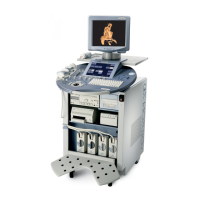
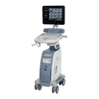
Details the system configuration and mechanical adjustments, including monitor, console, and trolley components.
Describes the basic system components and optional peripheral devices, detailing their modules and connections.
Explains the control center, digipots, flip switches, trackball, and navigation wheel functions.
Instructions on how to use the electronic user manual, including search functionalities and exiting the system.
Information on initial installation, first-time power-on, and recommended basic settings for the system.
Crucial warnings regarding temperature balance for condensation humidity and isolation transformer use for peripherals.
Step-by-step guide on powering on the system, including locating the standby switch and initial display.
Procedures for connecting and disconnecting probes, including cautions regarding freeze mode and cable handling.
Details on how to select connected probes, applications, and user programs for system adjustment.
Instructions on entering patient data for calculations, reports, and image identification, including worklist server use.
How to add text or auto annotations to ultrasound images, including cursor positioning and text entry.
Explanation of the [2D Mode] key function and how to adjust 2D settings via the sub-menu.
How to adjust the depth range of the ultrasound image, affecting frame rate and acoustic power indices.
Using the [Angle] digipot to select a part of interest, affecting frame rate due to smaller sector width.
Adjusting the overall brightness of the 2D image by controlling the gain amplification applied to received echoes.
Varying gain at specific depths to compensate for echo attenuation, affecting sensitivity and brightness.
Function to optimize contrast resolution based on scan area histogram, enhancing gray scale.
Controls acoustic output of the transducer, emphasizing minimum ALARA principle for power levels.
Selecting focal zones to optimize sharpness, with adjustments for number and depth of focal zones.
Adjusting frequency for resolution, penetration, and optimizing image quality for different tissue types.
Function to improve grayscale contrast and reduce artifacts, particularly useful for difficult-to-image patients.
Allows fine-tuning the system for different tissue types, adjusting parameter for adipose, solid, cystic or normal tissue.
Adjusts Volume O-Axis position in 2D mode, indicating acoustic block position and sweep direction.
Combines frequencies and focal ranges to improve penetration and resolution, reducing speckle and artifacts.
Increases scan area by steering ultrasound lines, enhancing field-of-view and frame rate.
Alternates image orientation (left/right, up/down) without rotating the scan head, indicated by an orientation marker.
Displays multiple 2D mode images simultaneously, with options for Single, Dual, and Quad screen layouts.
Stores scanned frames in cine memory for review, allowing image-by-image playback.
Magnifies the image in read- and write mode using the zoom control, with adjustable zoom factors.
Magnifies the 2D image in write mode, allowing placement and resizing of a zoom box for detailed viewing.
Accesses sub-menu functions for 2D mode, including Quality, Line Filter, Enhance, Dynamic Control, Persistence, and Reject.
Describes how to switch M mode on and use the M-cursor for time and motion echo information.
Details M mode operations including cursor position, activation, sweep speed, and gain control.
Explains M sub-menu functions such as Format, Enhance, Dynamic Control, and Reject for image adjustment.
Explains Pulsed Wave Doppler mode, including sample volume cursor, flow direction cursor, and spectral analysis display.
Details Continuous Wave Doppler mode, covering cursor position, activation, gain control, and sweep speed adjustments.
Describes activating CFM mode, the CFM box appearance, and gain control for the CFM mode.
Covers CFM operations like box position, size, gain control, quality, WMF, frequency, and threshold settings.
Details CFM sub-menu functions including Display Modes, Scale, Line Filter, Gently Color, and Artifact Suppression.
Explains activating PD mode, the PD box appearance, and key controls for quality, frequency, and PRF.
Covers PD operations like box position, size, gain control, quality, WMF, velocity range, and threshold.
Details PD sub-menu functions including Line Filter, Gently Color, Artifact Suppression, Ensemble, and Line Density.
Describes activating TD mode and the TD box appearance, noting that the key is only illuminated if the probe supports Tissue Mode.
Covers TD operations like box position, size, gain control, quality, velocity range, and inversion.
Details TD sub-menu functions including Scale, Line Filter, Gently Color, Baseline, Ensemble, Line Density, TD Map, Balance, Dynamic Set, and Smoothing.
Explains volume mode keys, 3D/4D pre-menus, and different possibilities for static 3D or real-time 4D acquisition.
Steps for acquiring 3D sectional planes, including activating volume mode, selecting visualization, user programs, and display formats.
Details the 3D/4D Submenu functions such as Render View Direction, 3D Gray Chroma Map, Utilities, Contrast, Background, Balance, and Power Threshold.
Guides on performing 3D rendering, including selecting visualization modes, user programs, and adjusting render box parameters.
Explains Real Time 4D acquisition principles, conditions, and operations, including volume box placement and quality selection.
Information on the VOCAL imaging program for virtual organ computer-aided analysis, including contour detection and volume calculation.
How to display the calculated (Color Angio) Histogram of the volume after calculation via the VOCAL program.
Defines how gray and chroma maps determine echo brightness and color tone, with options for 2D, M, D, and 3D imaging modes.
Explains how to graphically display gray scale or color distribution within a marked ROI, with options for 2D, 3D, and Volume histograms.
Instructions for displaying externally connected video signals (e.g., VCR) on the monitor and selecting video sources.
How to connect to the World Wide Web using the built-in browser, with trackball cursor assignment and exit procedures.
Information on displaying biopsy guidelines, programming biopsy lines, and safety precautions for biopsy procedures.
Allows selection of required Thermal Index (TIS, TIB, TIC) for display, with guidance on keeping index numbers low.
Explains how to switch on the Basic Measurement function, select measurement marks, and position/store measuring marks.
Details how to perform 2D and M distance measurements, including cursor positioning and fixing markers.
Describes methods for measuring circumference and area using Ellipse or Trace, including adjusting ellipse width.
Explains four methods for measuring structure volumes, including 3D MultiPlane measurement.
Covers methods for velocity measurement, including accelerated velocity, velocity ratio, and average velocity using trace methods.
Details measurements for specific applications, such as Hip Joint measurement and evaluation according to a table.
Provides tables showing system inaccuracies for distance, area, circumference, and volume measurements.
Explains how to switch on the Calculation function, select measurements using navigation wheel or keyboard shortcuts.
Details how to view, save/send, edit, transfer, and print patient reports, including OB, GYN, Cardiac, and Vascular reports.
Provides packages for OB calculations including Fetal Biometry, Fetal Long Bones, Fetal Cranium, AFI, and Fetal Doppler measurements.
Instructions on viewing OB patient reports, including graphs and history of each fetus, and exiting the report.
Covers GYN calculations for Uterus, Ovaries, Kidneys, Follicles, and Ovarian Arteries in 2D and Spectral-Doppler modes.
Steps to view GYN patient reports, including selecting pages and exiting the report.
Details cardiac calculations available in 2D, M Mode, Spectral-Doppler, and Color-Doppler modes for various heart structures.
Instructions on viewing cardiac patient reports, selecting pages, and exiting the report.
Explains vascular calculations for ICA, CCA, ECA, Peripherals, and Heart Rate, including 2D and Doppler modes.
Steps to view vascular patient reports, select pages, and exit the report.
Describes how to select, load, remove, back up, and transfer exams via the DICOM network.
Explains how to review images using View Mode, Exam Mode, Compare Mode, Layouts, and Full Screen options.
Details how to use various tools in Sonoview, including printing, exporting, reporting, measuring, and DICOM functions.
Information on printing images and reports, including printer connection and assignment of print keys.
Instructions for recording using the VCR, including remote control operations and remarks on source quality.
Procedures for storing scanned images or volumes in Sonoview or sending to an external DICOM server.
How to access the Setup desktop by selecting the [System] key in the "Utilities" menu.
Procedures for exiting the setup, either cancelling changes or saving and exiting.
Overview of the different pages available in the System Setup desktop, including General, User Settings, Peripherals, etc.
How to access the Setup desktop by selecting the [Measure] item in the "Utilities" menu.
Procedures for exiting the measurement setup menu, either by closing without saving or saving and exiting.
Lists the available pages in the Measurement Setup desktop: General, Obstetric, and Cardiac Measurement Setup.
Instructions on programming biopsy lines, including placing the probe in a water bath and positioning the needle.
Details the ergonomic design of probes for ease of use, connection, and handling, as well as cable design.
Precautions for handling probe cables to prevent damage, such as keeping them free from wheels and avoiding sharp bends.
Explains the use of the orientation marking on probes to identify the probe's side corresponding to the image orientation.
Information on probe labeling, including manufacturer, part number, serial number, and designation displayed on screen.
Lists probes and their intended applications across 2D, 3D/4D modes, detailing usage areas like Abdominal, Obstetrics, and Cardiology.
Overview of probe capabilities across different modes, including HI, FFC, CE, CW Doppler, and HPRF for various probe types.
Technical specifications for probes, including center image frequency and Doppler frequency ranges (Low, Mid, High).
Details on connecting, activating, deactivating, disconnecting, transporting, and storing probes, referring to specific chapters.
Instructions for inspecting probes and general environmental requirements for operation, storage, and transport.
Guidelines on handling precautions, watertightness, electrical shock hazards, and mechanical hazards related to ultrasound probes.
Advice on using protective sheaths to minimize disease transmission, including instructions and reordering information.
Information on risks of disease transmission, proper cleaning, disinfection, and use of sterile probe sheaths.
Introduces the four types of probes supported by the V730Pro system, detailing their designation prefixes.
Discusses concerns related to biopsy procedures, including patient preparation, mounting, and programming/display.
Basic concept of isolation transformer, IEC standards for interconnecting medical devices, and safety measures.
Lists various connectors for internal and external accessories, including main module, power supply, and monitor connections.
Crucial notes on leakage current limits, connecting equipment to the console, galvanic separation, and voltage settings.
Details the ECG module, patient connection cable, and its use for acquiring ECG signals for ultrasound image display.
How to switch ECG function on/off, alter displayed ECG strip, and use patient cables and electrodes.
Safety rules for operating the ECG preamplifier, including cable connections, intra-operative use, and potential signal influences.
Guidelines for handling electrodes and cables, referring to manufacturer's instructions for cleaning, maintenance, and repairs.
Function to insert an ECG line into the image display, adjust gain, velocity, position, and amplitude.
Details power requirements, consumption, noise level, mains outlets, EMC compliance, electrical, mechanical, and thermal safety.
Technical data for the transmitter, including frequency range, acoustic output control, focusing, and processing channels.
Receiver specifications covering frequency range, focusing, sample-rate, processing channels, and dynamic range.
Technical data for the scan converter, including video memory, image memory, gray scale values, depth range, and scan angle.
Information on cine loop memory capacity and call-up of sequences for manual and automated playback.
Lists available display modes for 2D, 3D, 4D, M Mode, Doppler, and Color Doppler scans, including image orientation.
Details signal processing parameters such as persistence filter, line filter, enhance, reject, gray scale dynamic, dynamic, and quality.
Information on patient data, clinic/doctor's name, and auto text memory capacity for system input.
Lists basic measurements, obstetric calculations, and cardiology calculations available within the system.
Details the capacity of user program memory for presets, applications, and probe settings.
Technical specifications for the Volume Scan Module, including scan size, lines, images, frame rate, rotation, and display modes.
Technical data for Spectral-Doppler, covering working modes, transmit frequencies, PRF, gain, WMF, and spectrum analyzer.
Technical data for Color-Doppler (CFM), including display modes, color coding steps, depth range, and CFM Map options.
Technical data for Tissue-Doppler (TD) mode, covering display modes, TD coding steps, depth range, and TD Map options.
Technical data for Power-Doppler (PD) mode, including display modes, PD window size, smoothing filter, and PD Map options.
Details the various interfaces available, including video, S-video, RGB, VGA, audio, footswitch, remote control, and net connection.
Specifications for the SVGA monitor, including type, resolution, scanning frequency, nominal current, and safety classification.
Information on optional external drives such as Magneto-Optical Drive and CD-RW drive, including capacity and environmental requirements.
Technical data for the MAN ECG preamplifier, including input, patient cable, frequency range, rejection filter, and safety tests.
| Category | Medical Equipment |
|---|---|
| Manufacturer | GE Healthcare |
| Model | Voluson 730Pro |
| Power Requirements | 100-240V AC, 50/60Hz |
| Imaging Modes | Color Doppler, Power Doppler |
| Applications | Obstetrics, Gynecology |
| Probe Connectors | Multiple probe connectors |
| Frequency Range | 2-10 MHz |