•
yes: continue with next step
7.
Select
Add to Report
to save the measurements to the current exam.
Follicle measurement method:
auto
1. Scan and freeze a follicle volume data set.
2. Select Volume Analysis and select SonoAVC™
follicle
.
3. Adjust the ROI.
4. Select
auto
and adjust the ROI shape if desired.
5. Start the measurement by selecting either
Left Ovary
or
Right Ovary
.
6. The rendered follicles and the result list are displayed on screen.
7. Edit the results if necessary.
8. Select
Add to Report
to save the measurements to the current exam.
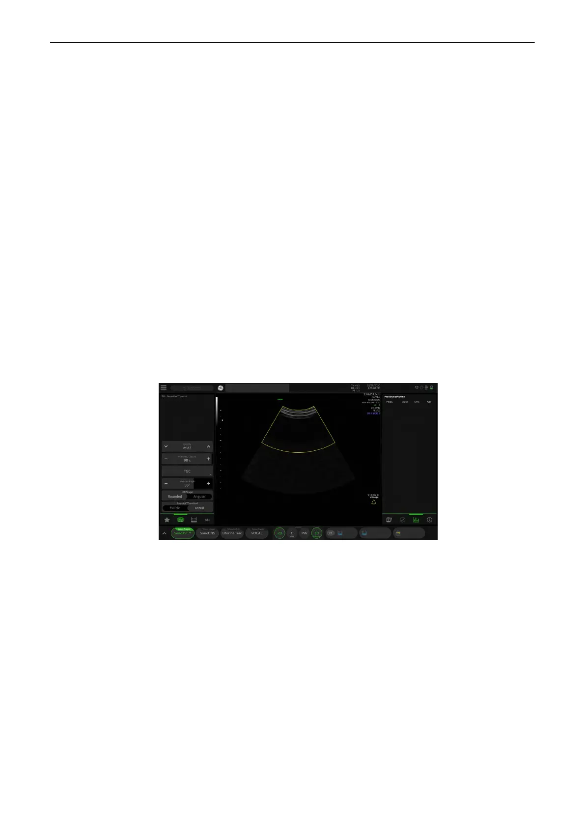
8.8.6.3 SonoAVC™antral2.0
Note
SonoAVC™antral
2.0
is an option.
Note
If a 4D Volume cine is present, the system will automatically switch to 3D Static when
SonoAVC™antral is pressed.
"Antral" means "antral follicle count". SonoAVC™
antral
2.0
enables to automatically detect and
count antral follicles within a ROI box in a 3D volume data set.
Volume Analysis Menu: SonoAVC™
antral
2.0
Figure 8-28 Volume Analysis Menu: SonoAVC™antral
2.0
ROI shape
Select the desired ROI shape:
•
angular
: the box has the shape of a rectangle
•
rounded
: the box has an elliptic shape with rounded corners that can be
adjusted by pressing (default position) and rotating (rounding the corners) the
rotary control. It is embedded in a rectangular box.
Start SonoAVC™
Select
Left Ovary
or
Right Ovary
to start SonoAVC™.
Ref. Image
Select the reference image to which all image dependent functions like parallel
shifts, rotations, etc. are applied.
3D and 4D Mode
8-42
Voluson™ SWIFT / Voluson SWIFT+ Instructions For Use
5831612-100 R
evision 4
 Loading...


