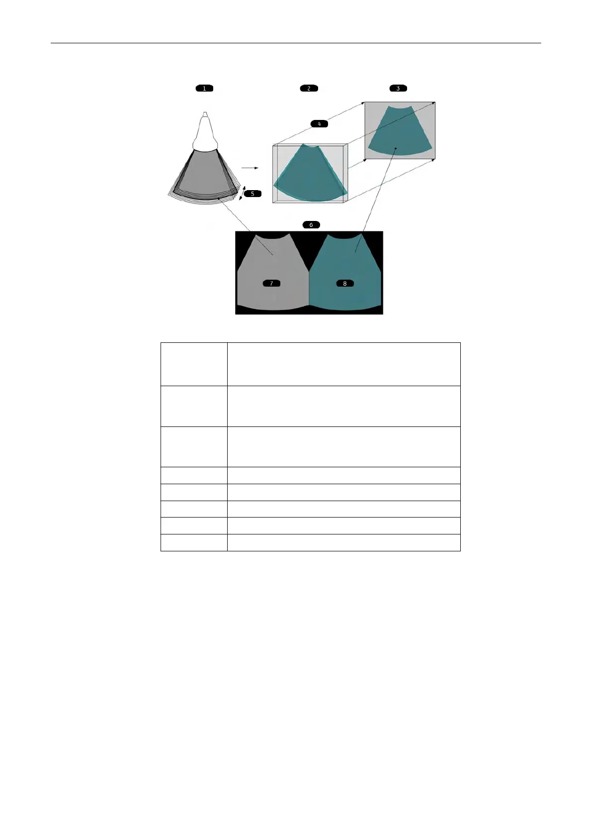
Figure 8-5 VCI-A principle
1 Scan Situation:
small 4D volume sweep
Vol. angle depends on slice thickness
2 Render Box:
Box size automatically derived from Vol. geometry. Box not
shown on screen.
3 Render result:
All single B-frames are rendered to one single VCI image (Thick
Slice Image).
4 Render direction
5 Vol. angle
6 Screen Display
7 Standard Image (center position of Vol. sweep)
8 VCI Image (Thick Slice rendered Image)
8.6.4 VCI OmniView
With help of OmniView, sectional planes derived from an entered trace can be visualized and
so special coronal planes are possible. The trace can be entered in the Vol. Pre image, or if a
volume data set, is present on image A, B or C. The trace can be a straight line, a curved line
or any freeform trace. Together with the VCI function images with less speckle pattern and a
highly improved tissue contrast can be archived.
3D and 4D Mode
Voluson™ SWIFT / Voluson SWIFT+ Instructions For Use
5831612-100 R
evision 4 8-13

 Loading...


