Do you have a question about the Leica DM RE and is the answer not in the manual?
Instructions for unpacking, assembly tools, and choosing an installation site.
Essential safety instructions for handling and setup.
Guidance on setting voltage and electrical safety measures.
Instructions for mounting deviating mirrors and lamp housings.
Instructions for changing halogen lamps in Lamphousing 106 and 106 z.
Critical safety information for handling Hg and Xe lamps, including explosion risk.
Advice on switching frequency, burner burn-in, and record keeping.
Emphasizes wearing safety clothing when assembling Xe burners due to explosion risk.
Proper insertion of burners, noting lettering orientation and seal points.
Specifications and features of mechanical stages 1187 and 1189.
Instructions for removing transit protection foam blocks from fixed stages.
Procedures for assembling and clamping interchangeable stages.
How to insert filter systems or reflectors into the incident light turret.
Components and steps for retrofitting the incident light axis.
Detailed steps for removing components and installing the incident light module.
Warnings regarding storing components and protecting optics from dust.
Description and main applications of the HC F diaphragm module.
Description and main applications of the HC RF diaphragm module.
Information on changing objective prisms and their configurations.
How to place or insert the polarizer for polarization contrast.
Details on different types of reflected light polarizers.
Description of the DM RD HC phototube as an automatic camera system.
Steps for turning on the microscope, setting selector switch, and adjusting brightness.
Individual setting of specimen clamp for Stage no. 1187 and 1189.
How to adjust the x-y drive for specimen positioning.
Instructions for loosening and adjusting the stage clamp for height setting.
How to use the mechanical dual knob drive for focusing.
Instructions and cautions for using the motorized focus system.
Steps for turning on the microscope and overview of motorized focusing controls.
Adjusting stage height using keystrokes and thresholds.
Setting stepwidths for fine focusing and using the focusing wheel.
How to set and delete upper and lower stage height thresholds.
How the coded nosepiece stores parameters for each objective.
How the coded nosepiece automatically calls up data and stores settings.
How the system protects against collisions and overloads.
Achieving perfect parfocality with mechanical and motor focus systems.
Important information and cautions before storing objective focus offsets.
Procedure for storing focus offsets using a specimen and calibration keys.
How to store objective magnification values during calibration.
Procedures for centering objectives for polarized light microscopy.
Two methods (I and II) for centering objectives accurately.
Steps for setting up transmitted light lamphousing 106 for illumination.
Setting up condensers and diaphragms for brightfield and Koehler illumination.
Adjusting condenser stops, diaphragms, and discs for brightfield.
Using phase contrast objectives and setting up illumination.
Centering light rings and phase rings for optimal phase contrast.
Setting up darkfield illumination with these condensers.
Using special darkfield condensers for improved darkfield imaging.
Adjusting light source, diaphragms, and condenser for polarization.
Step-by-step guide to crossing polarizers for accurate polarization contrast.
Details on index adjustment for IC/P polarizer and Analyser 360.
Accurate method using Bertrand lens or auxiliary telescope to cross polarizers.
Ensuring cleanliness and accuracy of polarizers and condensers.
Observing polarization effects and identifying birefringence.
How object brightness changes with stage rotation and identifying extinction positions.
Observing polarization effects and identifying birefringence.
Using adjustable compensators for exact phase difference measurements.
Essential steps for perfect ICT quality, including crossed polarizers.
Critical safety warnings for Hg and Xe lamps regarding explosion risk.
Setting up illumination for incident light brightfield microscopy.
Requirements and setup for incident light darkfield illumination.
Optimizing contrast with prism turret, aperture diaphragm, and compensators.
Setting polarizers and analyzers for transmitted light interference contrast.
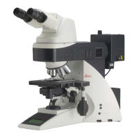
| Type | Upright Microscope |
|---|---|
| Focusing | Coaxial coarse and fine focusing |
| Optical System | Infinity-corrected optics |
| Condenser | Abbe condenser |
| Illumination | Halogen illumination |
| Eyepieces | 10x |
| Stage | Mechanical stage |
| Objective Lenses | Various objectives available (Plan, Achromat, etc.) |