g
Product Data Sheet – rev 4.2
July 4th 2003
Page 5
LightSpeed
With Xtream
CT Scanner System
GE Medical Systems-America
Milwaukee, USA - Fax: 1 414 544 3384
GE Medical Systems-Asia:
Tokyo, Japan - Fax: 81 425 85 5490
Hong Kong - Fax: 852 2559 3588
GE Medical Systems-Europe
Scan Fields of View
:
Biopsy: Simplified prescription for single
or multiple scans around an arbitrary table
position aids biopsy studies.
— 25 cm for adult head
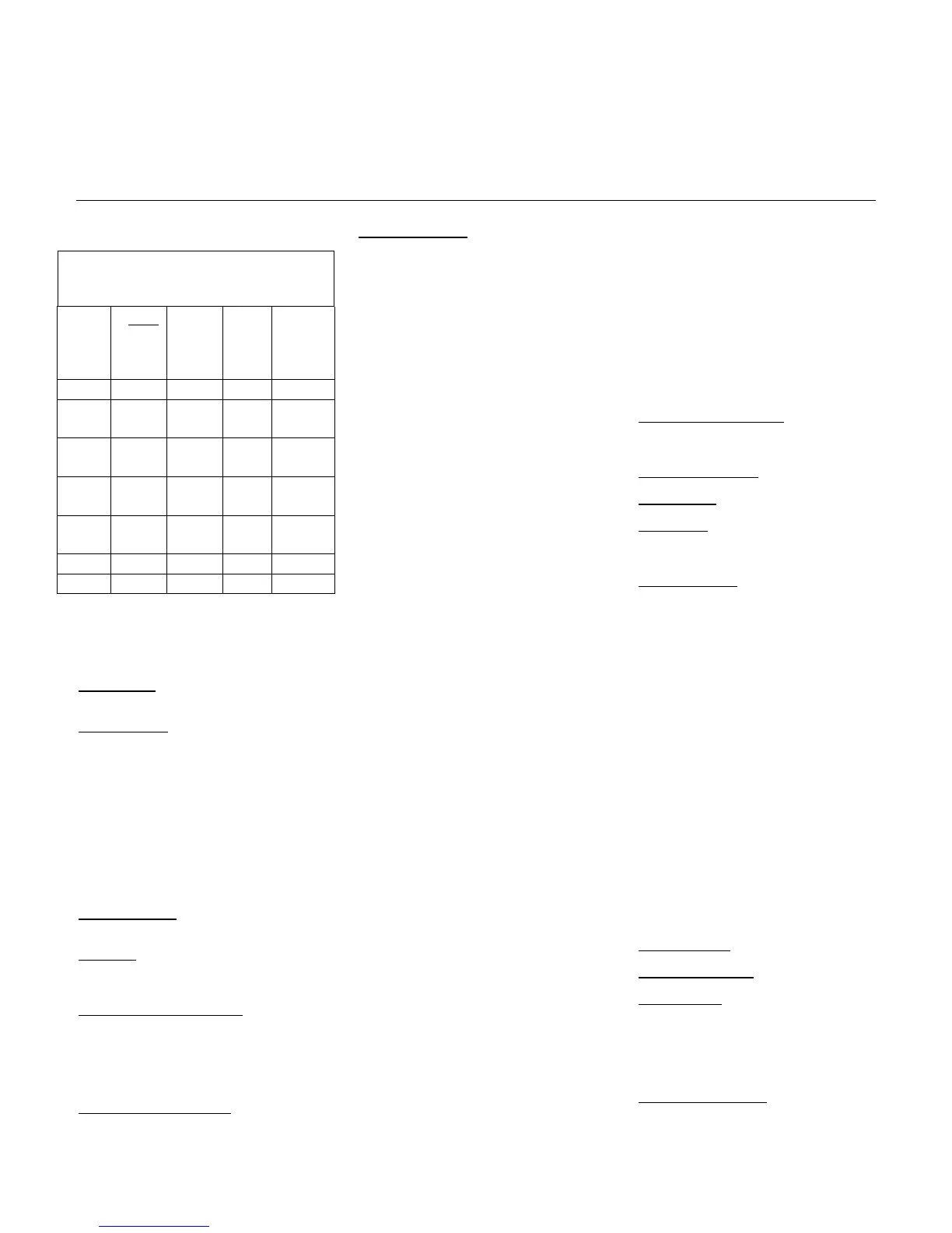
16-SLICE HELICAL MODES
Table Speed (mm/rotation)
Slice
Thick-
ness
(mm)
Pitch
0.5625
:1
0.9375
:1
1.375
:1
1. 75:1
0.625 5.625 9.375 13.75 17.5
1.25 5.625
11.25
9.375
18.75
13.75
27.5
17.5
35
2.5 5.625
11.25
9.375
18.75
13.75
27.5
17.5
35
3.75 5.625
11.25
9.375
18.75
13.75
27.5
17.5
35
5 5.625
11.25
9.375
18.75
13.75
27.5
17.5
35
7.5 11.25 18.75 27.5 35
10 11.25 18.75 27.5 35
— 25, 50 cm for body
Advanced Artifact Reduction (AAR)
Filter significantly reduces streaking
artifacts when highly absorbent objects
are in the field of view – ie: large
shoulders, screws.
— 25 cm for pediatric head
Helical Scan Enhancements
Helical Image Reconstruction
Full simultaneity allows complete image
display, processing and analysis, as well
as image archival and filming, concurrent
with scanning and reconstruction -- even
when acquiring helical images in a multi-
slice mode.
Reconstruction Algorithms: Soft Tissue,
Standard, Detail, Bone, Bone Plus, Lung
and Edge
Reconstruction Matrix
: 512
Confirm Rx to X-Rays on: < 15 sec. for
any state of tube and gantry; < 10 sec.
with the gantry rotating
Display Matrix
: 1024.
Display FOV
: Freely variable center/off-
center, prospective/retrospective target
selection.
Anatomical Programmer: a ten-region
anatomical selector allows quick and easy
access to 90 user-programmable
protocols per region. Separate selector
for adult and pediatric exams. There are
four selection tabs to select: GE, User,
Service and Most Recent Patient.
Copy/Paste is supported for easy
modification and copying of protocols.
CT Number Scale
: -1024 to 3071 HU
Helical Reconstruction Times:
Reconstruction time as fast 6
images per second
Helical Scan Parameters
Typical 0.167 sec. image-to-image
recon in normal 16 slice recon mode.
Ten user-defined regions. Each region
has one default protocol displayed with the
anatomical selector for very fast access to
most commonly used protocols
Scan Speeds: Full 360° rotational scans in
0.5, 0.6, 0.7, 0.8, 0.9, 1.0 seconds.
Maximum image-to-image cycle
time is ± 10% for prospective and
retrospective image-to-image display.
This applies for 512 matrix; any
display FOV; in AutoView (all
layouts); with concurrent filming and
image archival for all scan modes.
Scan Technique
:
Protocols include preset scan time, kVp,
mA, scan mode, slice thickness and
spacing, table speed, scan FOV, display
FOV and center, recon algorithm and
special image acquisition and processing
options
— kVp: 80, 100, 120, 140
— mA: 10 to 440 in 5 mA increments
— Power: 0.8 to 53.2 kW
Iterative bone processing
increases time by 250 milliseconds.
Iterative bone processing, which is
always enabled for adult head
scanning, reduces image artifacts in
head scans stemming from X-ray
beam hardening effects.
— Focal Spot Selection:
Any scan parameters may be edited for
each scan or all scans - either before or
during an exam. The number of scans
may also be easily changed.
Small spot for up to 24 kW
Large spot for greater than 24 kW
Single Acquisition
: 120 second scan
maximum.
AutoScan™: Fully automates longitudinal
table movement and start of each scan.
Minimum DFOV:
9.6 cm
Helical Tilt
: helical acquisition is possible
with the gantry tilted to a maximum of 30
degree, in half degree increments.
AutoVoice™: 3 preset (English) and 17
user-defined messages automatically
deliver patient breathing instructions;
especially useful for multiple helical
scanning.
Minimum Pixel Size:
0.19 mm
Queued Recon
: Requests will be
processed continuously and
simultaneously with other processes on
the system including scanning.
Prospective recon will be prioritized over
retrospective recon.
Multiple Acquisition Maximum
Scan Time:
Multiple scans may be acquired in one
series to produce up to 3,000 contiguous
helical images. Up to 2,881 seconds
helical coverage is possible in multiple
series.
Trauma Patient: Allows patient scans and
image display/analysis without entering
patient data before scanning.
Priority Recon Queuing
: One touch
selection marks most recent rotation for
Minimum Inter-Group Delay
(IGD): 5
seconds between adjacent helical scans
* Option
 Loading...