g
Product Data Sheet – rev 4.2
July 4th 2003
Page 12
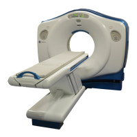
LightSpeed
With Xtream
CT Scanner System
GE Medical Systems-America
Milwaukee, USA - Fax: 1 414 544 3384
GE Medical Systems-Asia:
Tokyo, Japan - Fax: 81 425 85 5490
Hong Kong - Fax: 852 2559 3588
GE Medical Systems-Europe
do not support DICOM Print may require a
filming interface (purchased separately).
To save further filming cost, the Operator
Console can directly print to a postscript
printer such as the GE Color Printer available
as an option.
ImageWorks
ImageWorks software is designed to take
advantage of the Lightspeed
16
CT Scanner’s
computer and image processor. This desktop
environment includes image management and
networking.
Because some of the image analysis and
display features of ImageWorks replicate
those in Exam Rx, the next section describes
only features that are incremental or
significantly different.
Image Analysis
• Multi-Projection Volume
Reconstruction
(MPVR): Quick and easy
way to generate volumetric images for CT
angiography without thresholding data or
removing unwanted anatomy. An entire
volume is used to generate images in any
plane, creating real-time frames of
reference at the same time;
• Clinical utility is extended via two
additional modes:
− MIPS - enhances contrast and improves
visualization of calcifications
− Average - generates 2D radiographic
images;
•
Multi-planar Reformation (MPR):
Provides real-time assessment of anatomy
in offaxis planes. Sagittal, coronal, oblique
and curved planar reformations available;
•
Batch reformatting can also be defined
and executed for later viewing if desired;
• Image Addition and Subtraction: Includes
image addition of more than two images at
a time;
*Volume Viewer
Volume Viewer is an innovative and
powerfull suite of productivity enhanvers
(Volume Rendering, Volume Analysis and
Navigator) that includes :
•
Dynamic Volume Review™ for Fast
Screening
• Curved Volume Of Interest
• Protocol Management and Loading
• Review Layout Presets
• Multiple VR Objects Merge
• Pseudo Surface Shading Mode
• Predefined Cut Planes
• Volume Rendered Navigator views
• VR Preset save/recall
• 3D Rendered Lumen View
• Automatic Path Tracking
• Path Bridging (in case of occlusions)
• SmartCursor™ for Easy Navigation
• Synchronized Reformatted Views
• Cut visualization mode
*Advanced Vessel Analysis
Advanced Vessel Analysis is the ultimate
tool to assess and quantify vascular
structures, including stenosis analysis,
stent planning procedures, post stenting or
vascular surgery follow-up.
• protocol driven tools to perform quick,
flexible and accurate quantitative analysis of
vascular anatomy
• provides maximum, minimum and mean
intraluminal diameter measurements
• provides cross-sectional areas of true
orthogonal sections of the aortoiliac systems
at selected anatomical points
• clinical benefits include: stenosis sizing, pre-
and post- surgical assessment, stent
planning
• Measurements in % stenosis or mm of
stenosis, and measurement of length and
dimension of stenosis.
Image Display
• Magnifying Glass allows quick 2X mag
window that can be moved over an image.
• Image Scroll moves an image within its’ own
window.
• Groupings allow application of window/level
values, magnification/minification, image
scroll or flip and rotate to a user-defined
image set.
• Save State stores user-selected image
orientation and window/level with each data
set.
• Window/Level values may be:
• Preset to provide six on-screen instant
window/level settings
• Set independently for up to 16 images on
the screen
• User-modified in discrete or variable
steps
• Adjusted real-time on-image by holding
down the middle mouse button and
moving the mouse
• Cine mode provides paging in up to 4 view
ports of up to 128 previously-stored CT or
MR images at full selected display frame
rate. For more than 128 images, display
frame rate may be reduced.
• Cine mode also provides temporal, spatial
or manual playback loops.
• Text Page
Image Annotation
• Image annotation and cursor are shadowed
to permit ease in reading.
Image Management
• Images may be stored and retrieved via
Magnetic Optical Drive (MOD) media using
DICOM 3.0 format. This allows interchange
with other imaging systems supporting
DICOM 3.0 MOD media. Not all vendors
implementation of DICOM 3.0 are identical,
so please check with the manufacturer for
compatibility.
• Off-line retrieval of all image files. Images
may be viewed as soon as they are restored
from MOD.
•
Image retrieval time is approximately 30
images 512 per second.
VariViewer
TM
:
• VariViewer is an interactive axial review
mode that can change the slice thickness
reconstruction instantaneously.
• The user selects the volume to be
specifically analyzed and chooses the slice
thickness to be displayed as axial images.
The user can then save a number of recon
images sets, viewing a large number of
slices for pure axial review and filming.
* Option
 Loading...