Chapter 11 RESP Monitoring
11.1 Introduction
Measurement method: chest impedance. When the patient breathes, the thoracic activity causes a change in the thoracic
impedance between the two ECG electrodes. The monitor produces a respiratory wave on the screen by measuring the
impedance change (due to the movement of the thorax), then it calculates the respiration rate based on the waveform
cycle.
11.2 Safety information
WA RN IN G
n Respiratory measurement does not recognize the reason of suffocation, it will only give alarm if no next
respiration is checked within the predetermined time after the last breath, so it can not be used for
diagnostic purposes.
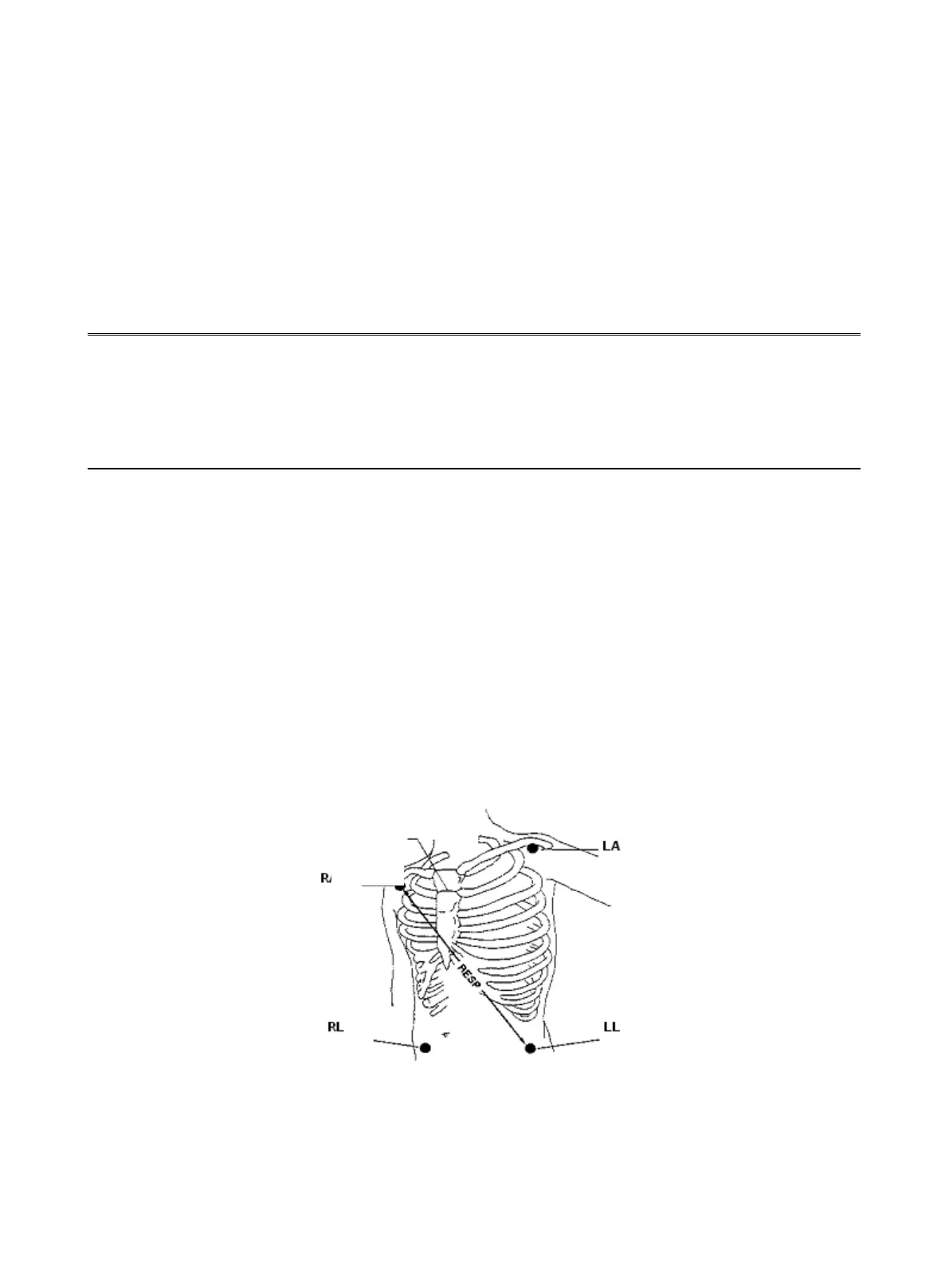
11.3 Placement for RESP electrode
As the skin is a bad conductor, in order to get a good respiration signal, process the skin where the electrode is placed is
necessary. See "ECG Monitoring" chapter for skin processing method.
For RESP monitoring, it is not necessary for additional electrodes, however, the electrode placement is important. Some
patients, due to their clinical condition, expand their chest laterally, causing a negative intrathoracic pressure. In these
cases it is better to place the two RESP electrodes laterally in the right axillary and left lateral chest areas at the
maximum point of breathing movement to optimize the respiratory waveform.
NOTE:
l The RESP monitoring is not recommended to be used on patients who are very active, as this can cause false
alarms.
Electrodes Placement (5-lead)
NOTE:
l Placing the red and white electrodes diagonally to obtain the optimal respiration waveform. Avoid the liver