Do you have a question about the Leica DM1000 and is the answer not in the manual?
Explains the meaning of symbols, pictograms, and text conventions used in the manual.
Provides general safety guidelines and compliance information for the microscope.
Details electrical specifications and safety precautions for microscope operation and power supply.
Provides instructions and regulations for the proper disposal of the microscope and accessories.
Specifies the recommended environmental conditions and location for microscope setup and operation.
Outlines procedures for safely disassembling and packaging the microscope for transport.
Details the steps for attaching and securing the microscope stage and specimen holder.
Explains how to adjust the focus stop to prevent objective collision and set focal planes.
Describes the procedure for mounting and using the stage lock mechanism.
Guides on how to properly insert and center the microscope condenser.
Instructions for mounting the tube and inserting eyepieces, including notes for fluorescence applications.
Provides guidelines for selecting, mounting, and arranging objectives on the nosepiece.
Details the process of replacing the tungsten halogen lamp in the transmitted light illumination system.
Explains the mounting procedure for the fluorescence illuminator before the tube.
Provides safety notes and instructions for inserting gas discharge lamps into the 106z lamp housing.
Describes the installation of the analyzer and polarizer components for contrast methods.
Details the steps for attaching the lambda plate compensator to the microscope base.
Discusses connecting a camera via adapter and provides a table for magnification calculations.
Explains how to use the ergomodule for raising the eye level of the tube opening.
Describes the base with adjuster wheels for optimizing the working position.
Details the optional magnification changer and its available magnification factors.
Guides on how to insert and use rechargeable batteries for the DM1000 LED microscope.
Explains how to connect the microscope stand and accessories to the external power supply.
Provides instructions on how to switch on the microscope and initial cautions for gas discharge lamps.
Details the procedure for setting up Köhler illumination for transmitted light brightfield illumination.
Explains how to check the centering of light rings for phase contrast objectives.
Guides on centering the 106z lamp housing and adjusting the arc image for proper illumination.
Instructions for switching on the microscope, particularly with gas discharge lamps.
Covers lengthening the coaxial pinion and adjusting the travel range of the stage.
Details how to change the side of the stage's coaxial pinion for right- or left-hand operation.
Explains the operation of coarse and fine focusing wheels and height adjustment of focusing wheels.
Discusses adjusting viewing distance, viewing angle, and beam splitting in photo tubes.
Provides guidance on using eyepieces, including inlaid reticles and vision correction.
Covers changing objectives, using immersion oil, and lockable immersion objectives.
Details how to adjust brightness for transmitted light and switch on fluorescence lamps.
Explains the function of the aperture diaphragm in controlling resolution, depth of field, and contrast.
Explains the role of the field diaphragm in protecting the specimen and improving contrast.
Details transmitted light contrast methods like brightfield, phase contrast, and darkfield.
Outlines the steps for setting up and using brightfield illumination with the microscope.
Guides on setting up and using phase contrast, including objective selection and condenser disk configuration.
Explains how to set up and use darkfield illumination with specific condensers and objectives.
Details how to achieve relief-like contrast using oblique illumination with specific condensers.
Describes the procedure for setting up and performing polarization microscopy, including polarizer and analyzer alignment.
Provides instructions for setting up and using the fluorescence illumination system.
Explains the required tools and calibration process for performing linear measurements with the microscope.
Details the method for measuring object thickness using stage height differences and refractive indices.
Describes the object marker tool used for scribing circles to mark objects during measurements.
Outlines the procedure for differentiating gout and pseudo gout crystals using the lambda plate compensator.
Lists common problems related to the microscope stand not responding and their possible causes/remedies.
Addresses issues with dark or unevenly illuminated images and flickering illumination.
Discusses problems with bringing the specimen into focus and their potential causes.
Troubleshoots issues with obtaining definite darkfield contrast.
Addresses problems related to achieving polarization contrast.
Troubleshoots issues where phase contrast is not possible due to specimen or setup issues.
Addresses problems with dark or weak fluorescence images.
Solves issues related to the decrease in the stage's travel range over time.
Provides guidance on using the dust cover to protect the microscope after use.
Details procedures for cleaning coated parts, glass surfaces, and objectives, including cautions.
Offers cautions for handling acids and bases to prevent contact with optics and mechanical parts.
Explains how to change fuses in the DM1000 stand and provides safety warnings.
Guides on how to equip the condenser disk with light rings, compensators, or lenses.
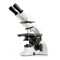
| Microscope Type | Upright Microscope |
|---|---|
| Focus | Coaxial coarse and fine focus knobs |
| Application | Research |
| Illumination | LED |
| Eyepiece | 10x |
| Stage | Mechanical stage |
| Condenser | Abbe condenser |
| Optical System | Infinity optical system |
| Magnification Range | 40x to 1000x |
| Nosepiece | 4- or 5-position nosepiece |