BiliChek Service Manual
6
1014988
2.0 Purpose of the Device
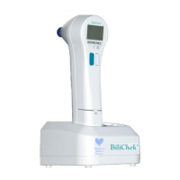
The BiliChek
®
Non-Invasive Bilirubin Analyzer accurately
determines bilirubin levels in newborn patients without a
blood sample regardless of their skin color, gestational age,
or post-natal age. This product provides rapid, point-of-care
bilirubin measurements as a replacement for traditional
clinical chemistry methods. These results are achieved with
no trauma to the patient, no risk of infection, and potentially
reduced cost of monitoring serum bilirubin by minimizing the
use of hospital personnel and supplies.
3.0 Theory of Operation
The BiliChek works by directing white light into the skin of the
newborn and measuring the intensity of the specific wavelengths
which are returned. By knowing the spectral properties of the
components within the skin, one can subtract out the interfering
components and determine the concentration of bilirubin.
Each photon has a characteristic wavelength. As light enters
skin tissue it can collide with the structural components such as
collagen fibers. When a collision occurs, the photon loses energy
and direction of travel is changed. This is called a scattering
event. If enough of these scattering events occur, the photon
completely loses its energy and is absorbed. If a photon is
scattered such that it is re-emitted from the skin, it is reflected.
Photons with longer wavelengths (in the red region of the
spectrum) are scattered less than photons with shorter
wavelengths (in the blue region of the spectrum). This
phenomenon is called wavelength-dependent scattering and
explains why the skin appears red when you shine a bright light
through it. It is also one of the reasons why the optical properties
of the newborns skin changes with advancing gestational and
post-natal age. As the skin matures, it becomes thicker and
there is greater keratinization of the cell membranes which
increases the scattering of incident light.
Photons of specific wavelengths are also preferentially absorbed
by certain molecules. By plotting the absorption against the
wavelength one can visualize characteristic absorption spectra
of the particular molecules. For example, melanin has a near-
linear absorption spectrum in the visible spectrum and, like the
scattering phenomenon, there is greater absorption of photons
with shorter wavelengths than in the red region of the spectrum.
Conversely, hemoglobin is a much more complicated absorber
which is compounded by the fact that oxyhemoglobin and
deoxyhemoglobin have different profiles. The peak absorption
of photons by bilirubin occurs at a wavelength of 460nm. This
is in the blue portion of the spectrum and is the reason why blue
lights are sometimes preferred for phototherapy. It is also in
the region of the spectrum at which hemoglobin absorption is
relatively low.
 Loading...