Do you have a question about the Siemens Uroskop ACCESS and is the answer not in the manual?
General safety guidelines for operating medical devices.
Lists required operating instructions for the TFT Monitor and camera.
Specifies necessary tools like a standard toolkit and oscilloscope for operation.
Details on connecting, adjusting, and image quality factors for the endoscopy camera.
Information on cable connections, power supply, and grounding for the endoscopy interface.
Explains monitor modes, image sources, and factory-set color and brightness parameters.
Troubleshooting for a dark TFT monitor, including connection checks.
Addresses monitor staying in X-ray mode by checking power and connections.
Solutions for monitor reverting to X-ray mode, focusing on signal path.
Diagnosing and fixing incorrect color display, checking RGB signal strength.
Describes visual distortions like smearing, tearing, and blooming.
Covers delay, skipping, and image sector size problems.
Smearing is an inherent characteristic not correctable by monitor adjustment.
Delays are due to image scanning and cannot be corrected by monitor adjustment.
Correcting tearing by adjusting camera signal or using a terminal resistor.
Skipping occurs during fast movement and is not monitor-adjustable.
Addresses causes of blooming related to camera, light source, and monitor settings.
Procedure for adjusting monitor contrast and brightness using test images.
Solutions for incorrect image sector size involve endoscope replacement or selection.
This document outlines troubleshooting procedures for the Endoscopy Option of the Siemens Uroskop ACCESS system. It covers general information, details about the endoscopy camera, interface, and monitor, and provides solutions for common video signal errors and image quality issues.
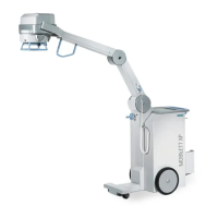
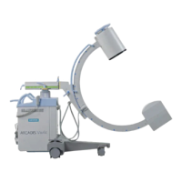
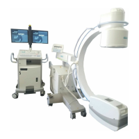
The Uroskop ACCESS Endoscopy Option integrates endoscopy video capabilities into the Uroskop ACCESS system. It allows for the display of endoscopic images on a Thin-Film Transistor (TFT) monitor, supporting various modes including X-ray, Ultrasound, and Endoscopy. The system is designed to process video signals from an endoscopy camera through a dedicated interface to the monitor. The endoscopy interface facilitates the transmission of video signals from the camera to the monitor, with a preference for S-Video input due to its superior signal quality. It also provides a 230 V supply voltage for the endoscope and includes grounding bolts for equipotential bonding, ensuring electrical safety. The system is capable of handling video signals from various manufacturers' endoscopy cameras, though optimal performance is achieved with cameras that adhere to a standard video signal of 1 Volt Peak/Peak (700 mV image signal; 300 mV synchronizing signal).
The Uroskop ACCESS Endoscopy Option offers flexible usage across different imaging modalities. The reference monitor can be switched between X-ray, Ultrasound, and Endoscopy modes via the control panel or system footswitch. In "Endo mode," images from the endoscopy camera intensifier are transmitted via the S-video input of the endoscopy interface. The system supports various endoscopes with different lenses and fields of view, allowing for adaptability to different examinations. Brightness, contrast, and background light settings for the reference monitor are factory-set for all modes, ensuring consistent image presentation. Additionally, four basic color settings are pre-configured, with options for X-ray reference (light and blue), neutral (for both X-ray and endoscopy), endoscopy-specific (red/brown), and a user-definable setting for individual customer requirements. The system is designed to display endoscopic images, and the quality of these images is dependent on the signal strength from the camera output.
Maintenance of the Uroskop ACCESS Endoscopy Option primarily involves troubleshooting and adjusting settings to ensure optimal image quality. The document provides detailed steps for addressing common video signal errors such as a dark TFT monitor, the monitor remaining in X-ray mode, switching back to X-ray mode, or displaying incorrect colors. These steps often involve checking video connections, measuring video signals with an oscilloscope, and verifying supply voltages.
For image quality issues like smearing, delay, tearing, skipping, blooming, and incorrect image sector size, specific corrective actions are outlined. Smearing and delay, being characteristics of digital cameras and monitors, are generally not correctable by monitor adjustment. Tearing, caused by incorrect synchronization, requires measuring the video signal at the endoscopy interface input and adjusting the camera to a 1 Volt Peak/Peak video signal. In exceptional cases, a terminal resistor can be connected to reduce the video signal.
Blooming, which manifests as overly bright or glaring images, can be addressed by reducing camera amplification, checking the light source controller and connection cables, or verifying the light regulator. Monitor-related blooming issues require checking and adjusting brightness and contrast settings using test images with chrome-plated objects. The document provides visual examples of correct and incorrect contrast settings to guide the user.
For incorrect image sector size, the recommendation is to replace obsolete endoscopes or use endoscopes provided by the camera manufacturer to ensure proper image alignment and sizing. The document also emphasizes the importance of adhering to general safety information for medical devices during any maintenance activities or tests. Regular checks of luminance using a test image and a Mavo Monitor are recommended, with monitor replacement suggested if maximum luminance falls below 100 cd/m².
| Category | Medical Equipment |
|---|---|
| Application | Urology |
| X-ray Generator | High-frequency generator |
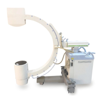
| Mobility | Mobile C-arm system |
| Imaging Technology | X-ray |
| Imaging Modality | X-ray |
| Power Supply | 50/60 Hz |
| Features | Digital image processing, dose reduction system |
| Dimensions | Varies by configuration |
| Spatial Resolution | High, varies with configuration |