Do you have a question about the Siemens SOMATOM Emotion and is the answer not in the manual?
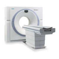
Specifies the purpose and application of the SOMATOM CT system for generating tomographic images.
Details the diagnostic and treatment preparation uses of the generated CT images by trained physicians.
Defines the qualifications and training required for personnel operating the SOMATOM CT system.
States that there are no known restrictions on the patient population for using the CT system.
Provides guidance on scanning pediatric patients and refers to external resources for more details.
Confirms that there are no specific situations known to contraindicate the use of the SOMATOM CT system.
Outlines essential qualifications and instructions for users to safely operate the CT scanner.
Lists available documentation, including Instructions for Use, Online Help, Quick Guide, and manuals.
Describes the scope and purpose of the Quick Guide, its limitations, and related documentation.
Details critical safety measures for preventing patient injury, radiation exposure, and equipment damage.
Guides users on the controlled restart procedure following an emergency stop of the system.
Outlines the steps to safely restart the CT system after a power interruption.
Explains how to manually retract the patient table top from the gantry during emergencies.
Describes the process of entering patient information into the system for examination setup.
Explains how to choose predefined scan protocols to set up the examination procedure.
Provides instructions and safety precautions for correctly positioning the patient on the table.
Details the first step of a CT examination: acquiring a topogram for planning purposes.
Guides users on defining the anatomical region for scanning using the topogram.
Explains how to use Automatic Patient Instruction recordings to guide patients during scans.
Describes the process of acquiring cross-sectional images (tomograms) for diagnosis.
Explains how to create axial images from scan data using reconstruction jobs.
Details the process of creating 3D image reconstructions for improved workflow efficiency.
Covers the final steps to close a patient examination and save the acquired images.
Describes how to load and navigate through patient image series using the browser function.
Explains how to adjust image contrast and brightness to optimize image display.
Details the procedure for magnifying or reducing the size of images for closer inspection.
Describes how to move zoomed images within the display segment for better viewing.
Explains how to mark regions of interest and evaluate statistical gray scale values in images.
Guides users on measuring distances between points on CT images.
Details the process of measuring angles between lines on CT images.
Provides instructions on how to select and save image series from the viewing session.
Explains how to load image series into the 3D task card for postprocessing.
Guides users on adjusting the orientation of image segments for better visualization.
Describes how to find and view specific slices within the 3D dataset.
Details the procedure for generating custom oblique views from the image data.
Explains how to create a series of parallel images from a selected range.
Details the process of printing images onto film using a connected camera.
Provides instructions for saving examination data onto approved DVD media.
Explains how to transfer patient data between systems using a USB device.
Guides users on sending examination data to other workstations over a network.
Describes the procedure for removing patient data from the system database.
| Type | CT Scanner |
|---|---|
| Gantry aperture | 70 cm |
| Tube voltage | 80, 110, 130 kV |
| Maximum scan range | 200 cm |
| Maximum Scan Field of View | 50 cm |
| Generator Power | 32 kW |
| Image Matrix | 512 x 512 |
| Rotation time | 0.8 s, 1.0 s, 1.5 s, 2.0 s (depending on configuration) |
| Patient weight capacity | 200 kg |
| Table Weight Limit | 200 kg |
| Image Reconstruction Time | Up to 8 images/second |