Do you have a question about the Sirona galileos comfort plus and is the answer not in the manual?
Provides contact details for technical queries and manufacturer information.
Specifies the product's intended applications and contraindications for medical use.
Explains the manual's organization, danger levels, and used formats/symbols.
Covers unit markings, ventilation, condensation, and basic safety principles.
Defines requirements for personnel operating the system safely and effectively.
Details procedures and equipment for minimizing radiation exposure during operation.
Explains the emergency stop function and safe use of laser light localizers.
Covers hygienic practices and safe operation of the touchscreen interface.
Specifies conditions and requirements for ensuring the unit operates without malfunctions.
Addresses EMC compliance and risks from electromagnetic fields and interference.
Details ESD risks, protective measures, and the physics of static charges.
Presents comprehensive technical specifications of the GALILEOS system components.
Illustrates technical aspects with cooling curves, beam angles, and anode details.
Provides measured values for secondary scattered radiation at different angles.
Lists certifications and compliance with relevant international standards (IEC).
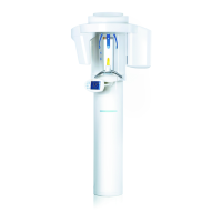
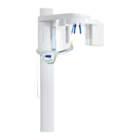
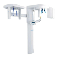
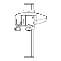
Identifies and describes the operating and display elements on the main GALILEOS unit.
Details the controls and adjustments for the head fixation device.
Explains the layout and individual control elements of the Easypad touchscreen.
Covers general functions like color codes, symbols, program settings, and service menus.
Details how to access and adjust program settings in the second program level.
Describes how to adjust touchscreen settings like click tone and intensity.
Lists and describes various bite blocks, supports, and fasteners for patient positioning.
Details accessories for the head fixation device, including ear olives and forehead pads.
Explains the use and types of hygienic protective sleeves for maintaining cleanliness.
Describes the test phantom used for acceptance and constancy testing of the unit.
Details the VO5 program for high-resolution scans and 3D data set creation.
Explains the High Contrast (HC) mode for improved display of hard structures.
Explains the High Definition (HD) mode for improved visualization of structures.
Describes the VOG program for scanning small objects like dental templates.
Covers steps before exposure, including fitting accessories and powering on the unit.
Details how to correctly attach and secure various accessories like bite blocks and holders.
Provides instructions for safely powering on the X-ray unit and system checks.
Explains the initial display on the Easypad touchscreen after system startup.
Guides on choosing kV/mAs/dose values based on patient size and program.
Details how to adjust the mechanical diaphragm for different jaw views.
Details methods for positioning patients with bite blocks, head fixation, and spherical blocks.
Step-by-step guide for positioning patients using standard and chin rest bite blocks.
Instructions for positioning patients, particularly for orthodontics, using the head fixation device.
Guidance on using the spherical bite block for patient positioning and implant templates.
Explains the process of initiating and completing an X-ray exposure using the release button.
Describes how to use the remote control for releasing exposures and system control.
Provides a reference for system messages and required actions.
Explains the format of error codes (Ex yy zz) and their classifications.
Details causes and solutions for a specific system error message.
Instructs on proper cleaning and disinfection of the unit using approved agents.
Covers the process and requirements for sterilizing compatible accessories.
Outlines required inspections and preventive maintenance schedules.
Procedures for safely dismantling and reinstalling the X-ray unit.
Information on proper disposal of electrical devices, including the X-ray tube.
Presents radiation exposure data in terms of DAP for various programs and patient types.
Provides effective dose values (Deff) for different imaging modes and patient parameters.
| Model | GALILEOS Comfort Plus |
|---|---|
| Type | Cone Beam Computed Tomography (CBCT) |
| Manufacturer | Sirona Dental Systems |
| Scan Time | 14 seconds |
| Image Resolution | Up to 0.125 mm |
| X-ray Tube Voltage | 85 kV |
| Power Requirements | 230V, 50/60 Hz |
| Operating System | Windows |
| Field of View (FOV) | 15 cm x 15 cm (standard), adjustable |