Do you have a question about the Carestream DENTAL CS 9600 and is the answer not in the manual?
Describes the alphanumeric digital soft-touch console for interacting with panoramic and 3D functions.
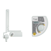
Explains how to launch radiological image acquisition using the remote control outside the X-ray room.
States the need to check computer system configuration compatibility with CS 9600 software.
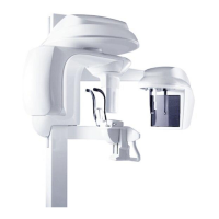
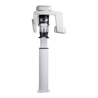
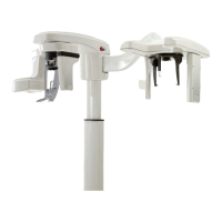
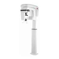
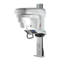
Describes the user-friendly interface for CS 9600, featuring Panoramic and 3D acquisition.
Details the interface for 2D imaging acquisition functions of the CS 9600.
Describes the interface for 3D imaging acquisition functions of the CS 9600.
Step-by-step guide to powering on the CS 9600 unit and workstation.
Details the process for powering on the CS 9600 workstation.
Guides users on how to access the Acquisition interface via CS Imaging or DICOM Worklist.
Details features and instructions for safer use of the CS 9600 with pediatric patients.
Explains patient size icons and their relation to reduced exposure values for children and adolescents.
Recommends using low-dose protocols for diagnostic tasks requiring lower resolution for pediatric patients.
Discusses reducing the Field of View for 3D imaging on children to lower radiation dose.
Informs that estimated X-ray dose emission is displayed during acquisition to evaluate risk vs. benefit.
Covers various panoramic radiological exams like full, segmented, bitewing, and TMJ images.
Covers panoramic exams for Maxillary Sinus and Sinus AP, PA, and Lateral views.
Guides on selecting program, FoV, and patient type for 3D wrist imaging.
Procedure for launching the X-ray acquisition for 3D wrist imaging.
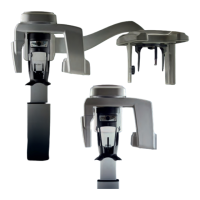
Instructions for preparing and positioning patients for 3D face scan imaging.
| Patient Positioning | Seated or standing |
|---|---|
| X-ray Tube Voltage | 60 - 90 kV |
| Software Compatibility | CS Imaging Software |
| Technology | CBCT |
| 3D Dose | Low-dose protocols available |
| Imaging Modalities | CBCT, Panoramic, Cephalometric |
| Voxel Size | 75 μm to 400 μm |
| Scan Time | 5-20 seconds |
| X-ray Tube Current | 3 - 10 mA |
| Detector Type | Flat Panel Detector |
| Dimensions | Varies depending on configuration. Refer to manufacturer's documentation. |
| Weight | Varies depending on configuration. Refer to manufacturer's documentation. |