20
Average measurement may be used to
locate the posterior reference points
whenever you do not vary the vertical
dimension of the casts on the articula-
tor, or, in other words, when the
mandibular cast is to be transferred to
the articulator by means of an interoc-
clusal record taken at the correct verti-
cal dimension and the vertical dimen-
sion is not going to be changed on the
articulator.
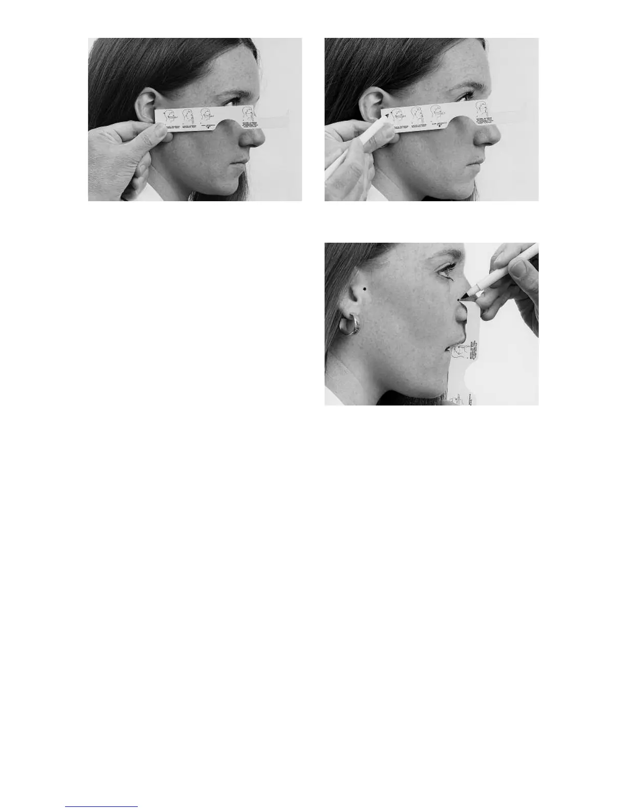
Place the “reference plane locator”
along the right side of your patient’s
face. It should extend from the middle of
the upper border of the external audito-
ry meatus to the “outer canthus” of the
eye. In other words, the reference plane
locator should extend from the middle
of the upper border of the ear-hole to
the outer corner of the eye (fig. 33).
There is a small hole in the upper poste-
rior area of the locator. Once the locator
is in position on the patient’s face, use
your felt-tipped pen to gently mark
through the hole onto the face (fig. 34).
Make the mark on both sides of the
patient’s face.
The position of the “anterior reference
point” is measured up 43 millimeters
from the “incisal edges” of the central or
lateral incisor, toward the inner corner of
the eye. The notched out area of the
“reference plane locator” is used to
make this measurement. The notch is 43
millimeters in length.
Simply rest the lower edge of the notch
on the incisal edge of the right central or
lateral incisor. On an edentulous patient
measure up from the low lip line. The
“low lip line” is the lower border of the
upper lip when it is in repose. In either
case, mark the anterior reference point
below the inner canthus of the right eye
where the top point of the locator touch-
es the patient’s face (fig. 35).
fig. 33 fig. 34
fig. 35