21
M
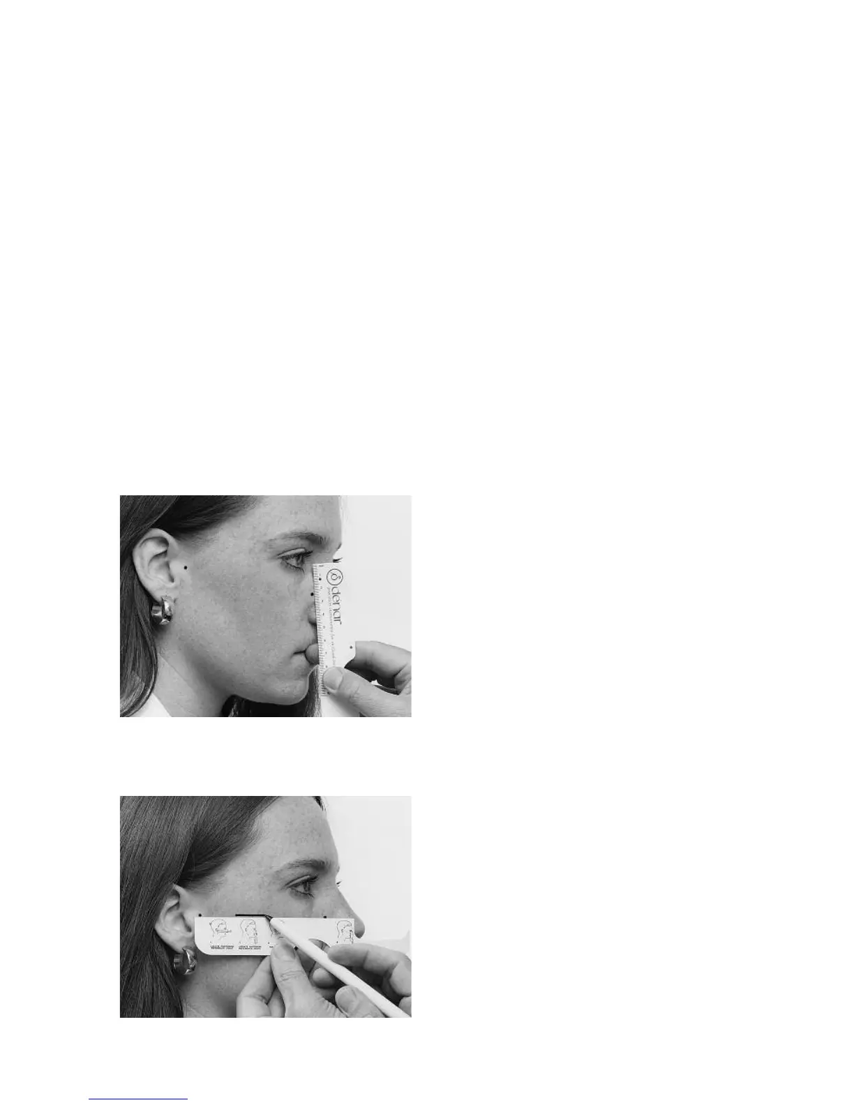
easure the distance between the ante-
rior reference point and the inner can-
thus of the eye (fig. 36). Record this
measurement in the patient’s file for
future reference. In this way, if the ante-
rior teeth are removed or modified the
same anterior reference point can be
located by measuring downward from
the fixed immovable inner canthus of
the eye.
The final step is to mark the “horizontal
reference plane” on the right side of the
patient’s face. Just line the ruler up
between the anterior and posterior ref-
erence points. Hold the ruler so that it is
just out of contact with the patient's
skin, so that it will not displace the skin,
and then draw a short line on the side of
the face. This line represents the “hori-
zontal reference plane” (fig. 37).
Y
ou will therefore notice that the hori-
zontal reference plane is identified on
the face of the patient by two posterior
reference points in the area of the termi-
nal hinge axis and one anterior reference
point located 43 millimeters above the
incisal edges of the maxillary anterior
teeth or low lip line of the patient.
MAKING THE FACEBOW/
EARBOW REGISTRATION
(Assembling the Facebow/Earbow
on the patient)
The components of the kit needed are:
the bitefork, anterior crossbar, reference
rod, reference rod clamp, and the right
and left facebow side arms with nylon
earplugs at the ends of the posterior ref-
erence slides (fig. 31).
Attach the bite fork to the crossbar so
the reference rod clamp is to the
patient’s right, and the u-shaped part in
the bite fork is above the crossbar (fig.
38). Then load the upper surface of the
bite fork with two thicknesses of base-
plate wax (fig. 39). Soften the wax to a
dead soft consistency in warm water or
an open flame, and then put the loaded
bite fork in the patient’s mouth to get a
light indexing impression of the maxil-
lary teeth. When the bite fork is first
placed in the mouth, be certain to line
up the crossbar so that it is parallel to
the coronal and horizontal planes of the
patient. Also be sure to be careful not to
depress or displace any mobile teeth
__
all you really need is a slight impression
of the tips of the cusps (fig. 40).
Remove the bite fork from the patient’s
mouth, and place the maxillary cast, if
available, in the bite fork to confirm
accurate seating. If the maxillary cast
seats accurately in the bite fork, you can
now begin assembly of the facebow
record.
Put the bite fork assembly back in the
patient’s mouth, indexing it to the maxil-
lary teeth. Have the patient hold the bite
fig. 36
fig. 37